Single photon emission computed tomography developer and preparation method thereof
A technology of single photon emission and tomography, which is applied in the preparation methods of peptides, preparation of X-ray contrast agents, chemical instruments and methods, etc., can solve problems such as difficult targeting, high preparation difficulty, and low cell uptake rate
- Summary
- Abstract
- Description
- Claims
- Application Information
AI Technical Summary
Problems solved by technology
Method used
Image
Examples
Embodiment 1
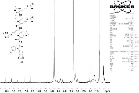
[0040] Tyrosine-cysteine-arginine-arginine-valine-arginine and valine-arginine-cysteine-arginine-tyrosine were firstly synthesized by solid-phase synthesis Amino acid-arginine two peptides were synthesized separately.
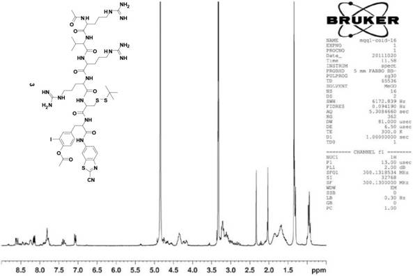
[0041] The second compound X in this example 2 The synthetic route of (Acetyl-Arg-Val-Arg-Arg-Cys(StBu)-Tyr(I-125)-CBT) is as follows:
[0042]
[0043] First, the tyrosine-cysteine-arginine-arginine-valine-arginine peptide was synthesized by solid-phase synthesis, and the specific steps were as follows: 0.33 mmoles of 2-chlorotrityl After the base chloride resin was swelled in 2 ml of N,N-dimethylformamide for five minutes, 0.66 mmol of the first amino acid was added to the reactor Add 0.66 mmol N,N-diisopropylethylamine, react for two hours, react with 30 microliters of methanol for 20 minutes, cut off the first amino acid protecting group, Kaiser test shows blue, add activated 0.55 Millimoles of the second amino acid cysteine reacted for two hours, K...
Embodiment 2
[0066] Embodiment 2: in vitro verification experiment
[0067] What adopted in the in vitro experiment of this embodiment is the third kind of compound X 3 At a concentration of 100 μmol / L, in a concentration of 100 mmol / L 4-hydroxyethylpiperazineethanesulfonic acid buffer, 1 mmol / L trichloroethyl phosphate, 1 mmol / L calcium chloride, and 5 μL of furin, the total volume was In 50 μL of the solution, incubate at 30°C for 16 hours to condense into dimers and self-assemble into nanoparticles.
[0068] Figure 5 The third compound X in this example is given 3 Transmission electron microscopy characterization of in vitro condensation-formed nanoparticles; Figure 6 It is the third compound X in this example 3 Dynamic light scattering analysis of in vitro condensation to form nanoparticles; Figure 7 It is the third compound X in Example 2 3 UV absorption at 500-700nm before and after in vitro digestion.
[0069] in Figure 5 It is a transmission electron microscope (TEM) ch...
Embodiment 3
[0070] Embodiment 3: cell uptake experiment
[0071] Firstly, the malignant breast cancer cell line MDA-MB-468 was cultivated, and the specific operation was as follows: the malignant breast cancer cell line MDA-MB-468 was cultured in DMEM medium containing 10% bovine serum albumin by volume concentration, and the cells were cultured in a volume concentration of 5% %CO 2 Cultured in a 37°C incubator in an air environment, and the medium was changed every other day.
[0072] Then do the cell uptake experiment, the specific operation is as follows: the malignant breast cancer cell line MDA-MB-468 is planted in a six-well plate, with about 1 million cells in each well, and the volume is 1mL. 5% CO 2 After six hours of incubation in a constant temperature incubator at 37°C in an air environment, three of the wells were replaced with 1 μCi of the second compound X 2 of fresh medium and the other three wells were replaced with 1 μCi of the sixth compound Y 2 Then collect part of...
PUM
| Property | Measurement | Unit |
|---|---|---|
| size | aaaaa | aaaaa |
| size | aaaaa | aaaaa |
Abstract
Description
Claims
Application Information
 - R&D
- Intellectual Property
- Life Sciences
- Materials
- Tech Scout
- Unparalleled Data Quality
- Higher Quality Content
- 60% Fewer Hallucinations
Browse by: Latest US Patents, China's latest patents, Technical Efficacy Thesaurus, Application Domain, Technology Topic, Popular Technical Reports.
© 2025 PatSnap. All rights reserved.Legal|Privacy policy|Modern Slavery Act Transparency Statement|Sitemap|About US| Contact US: help@patsnap.com


