Application of 5-hydroxymethylcytosine and method for detecting distribution of 5-hydroxymethylcytosine in mouse embryo

A technology of hydroxymethylcytosine, mouse embryos, applied in the field of biology

Inactive Publication Date: 2012-10-31
SHANGHAI INST OF BIOLOGICAL SCI CHINESE ACAD OF SCI
View PDF1 Cites 3 Cited by
  • Summary
  • Abstract
  • Description
  • Claims
  • Application Information

AI Technical Summary

Problems solved by technology

In addition, in addition to the DNA repair process, another possible mechanism for the demethylation of 5mc is the oxidation of the methyl group, followed by a series of continuous secondary reactions that eventually lead to the regeneration of cytosine

Method used


Image

  • Application of 5-hydroxymethylcytosine and method for detecting distribution of 5-hydroxymethylcytosine in mouse embryo
  • Application of 5-hydroxymethylcytosine and method for detecting distribution of 5-hydroxymethylcytosine in mouse embryo
  • Application of 5-hydroxymethylcytosine and method for detecting distribution of 5-hydroxymethylcytosine in mouse embryo

Examples


Embodiment 1

[0033] Example 1 Extraction and processing of mouse embryos

[0034] Select 4-5 weeks old female mice with closed vaginal opening as donors, and inject PMSG (7.5 IU) into each mouse intraperitoneally around 3:00 pm.

[0035] After 47 to 48 hours, each mouse was intraperitoneally injected with HCG (7.5 IU), and housed with normal male mice. The female rats were observed before 9:00 am the next day, and those with vaginal suppositories took them out for use.

[0036] At around 10:30, the female rats were killed by neck dislocation, the oviducts were taken out in M2 culture medium, the ampulla of the oviducts was torn open with forceps, and the fertilized eggs flowed out together with the granulosa cells. Digest with hyaluronidase, suck out the fertilized eggs, wash them in M2 solution, and finally put them in M16 solution with 5% CO 2 , 37 ° C incubator culture.

Embodiment 2

[0037] Example 2 In vitro fertilization of mice

[0038] Select 4-5 weeks old female mice with closed vaginal opening as donors, and inject PMSG (7.5IU) intraperitoneally into each mouse.

[0039] After 47-48 hours, each mouse was intraperitoneally injected with HCG (7.5 IU).

[0040] 12 to 13 hours after the injection of HCG, take a mature male mouse and kill it by breaking its neck. Take its epididymis tail, collect its sperm under a microscope, and place it in the pre-balanced HTF culture medium in 5% CO. 2 , 37°C incubator for 1 hour.

[0041] Eggs were taken from the female mice (the method was the same as above), and placed in the pre-balanced HTF culture medium. After the sperm capacitation is completed, an appropriate amount of sperm is taken to fertilize the egg.

[0042] After 4-6 hours of fertilization, the eggs were washed in freshly extracted and balanced HTF medium, and continued to be cultured in the incubator. A part of eggs are taken out at different set t...

Embodiment 3

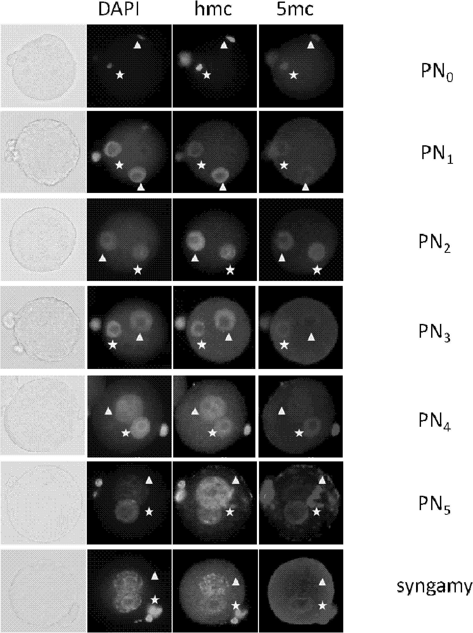
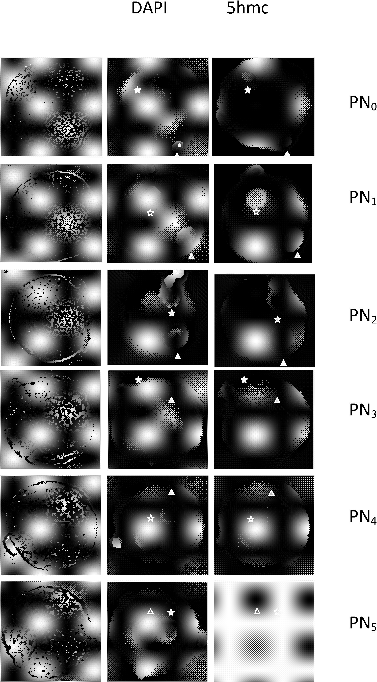
[0043] Example 3 Immunofluorescence staining of mouse embryos

[0044] The desired embryos were fixed in 4% PDF solution for 15 minutes at room temperature.

[0045] Wash in 1X PBS solution and treat with 0.2% Triton X-100 for 15 minutes at room temperature.

[0046]Wash in 1X PBS solution (PBST) containing 0.05% Tween20, and treat with 2N HCl for 30 minutes at room temperature, then immediately treat with 0.1M Tris-HCl (pH=8.0) for 15 minutes

[0047] Washing was performed in PBST solution, followed by blocking (block) for 1 hour in PBST solution containing 1% BSA.

[0048] The embryos were transferred into the anti-5mc / 5hmc antibody (diluted with blocking solution), placed at 37°C for 0.5-1 hour, and the positive signal was observed by fluorescent antibody.


PUM


Abstract

The invention discloses application of 5-hydroxymethylcytosine and a method for detecting distribution of 5-hydroxymethylcytosine in mouse embryo. The detection method comprises the following steps: (a) allowing a mouse to perform natural insemination or in vitro fertilization, and extracting the mouse embryo; (b) dyeing the mouse embryo; and (c) observing staining by virtue of a microscope or performing quantitative analysis. By adopting the detection method, the distribution of 5hmc in a mouse pre-implanted embryo is analyzed, the dynamical changing situation of 5mc and 5hmc in paternal and maternal chromosomes in the mouse pre-implanted embryo developing phase can be proved, and the important role of 5hmc to epigenetic reprogramming process in the mouse pre-implanted embryo developing phase can be disclosed, so that a new prospect is opened for studying a DNA (deoxyribonucleic acid) demethylation mechanism, and the 5-hydroxymethylcytosine can be used as a male nucleus marker or a female nucleus marker.

Description

technical field [0001] The invention belongs to the biological field and relates to the application of 5-hydroxymethylcytosine and a detection method for its distribution in mouse embryos. Background technique [0002] Methylation of 5-cytosine is an important part of the epigenetic code. Cellular differentiation, X-chromosome inactivation, recombination, and malignant transformation are major events in which significant epigenetic changes occur, including remodeling of DNA methylation patterns. Despite the relatively stable and heritable nature of DNA methylation in somatic cells, genome-wide DNA demethylation occurs in both developing primordial germ cells and fertilized eggs (zygotes). In zygotes, the demethylation of the markedly asymmetric DNA of the paternal and maternal genomes appears to occur in the same oocyte cytoplasm, starting as early as 6 hours after fertilization, when the paternal genome undergoes active DNA demethylation without significant demethylation ...

Claims


Application Information

Patent Timeline
Patent Type & Authority Applications(China)
IPC IPC(8): G01N33/58G01N1/28G01N1/30
Inventor 陈蘋贾微
Owner SHANGHAI INST OF BIOLOGICAL SCI CHINESE ACAD OF SCI
Why Eureka
Try Eureka