Extraction method of mouse liver sinusoidal endothelial cells

A technology of endothelial cells and extraction methods, which is applied in the field of extraction of mouse liver cells, can solve the problems of insufficient purity and poor cell viability, and achieve the stable effect of high cell purity, low cost, complete protection and activity

Inactive Publication Date: 2012-11-21
王慧 +2
View PDF0 Cites 6 Cited by
  • Summary
  • Abstract
  • Description
  • Claims
  • Application Information

AI Technical Summary

Problems solved by technology

[0005] The purpose of the present invention is to make up for the defects of the prior art, which are easy to produce mechanical damage when separating mouse liver sinusoidal endothelial cells, poor cell viability, state decline, and insufficient purity caused by chemical pr

Method used


Image

  • Extraction method of mouse liver sinusoidal endothelial cells
  • Extraction method of mouse liver sinusoidal endothelial cells
  • Extraction method of mouse liver sinusoidal endothelial cells

Examples


Embodiment

[0034] The steps for extracting hepatic sinusoidal endothelial cells from mice are as follows:

[0035] 1) Select normal male KM mice, about 9-10 weeks old, weighing between 20-25g, and inject 0.1-0.2ml of gadolinium chloride solution (about 10mg-20mg / 25g) into the tail vein for two consecutive days before surgery. Fasting the day before, no water restriction;

[0036] 2) Disinfect instruments and animal workbenches, pre-irradiate the experimental bench with ultraviolet light for 1 hour, pre-warm D-hanks at 37°C, 1640 culture medium, and collagenase IV solution with a mass concentration of 0.05%;

[0037] 3) Take a mouse for intraperitoneal injection of 0.1ml of pentobarbital sodium solution with a mass concentration of 1% (10mg / ml), about 40mg / kg, and 0.2ml of heparin sodium solution with a concentration of 200U / ml, about 40U / ml only;

[0038] 4) After the mice were completely anesthetized, they were fixed on the animal operating table, and the abdomen of the mice was disin...

experiment example 1

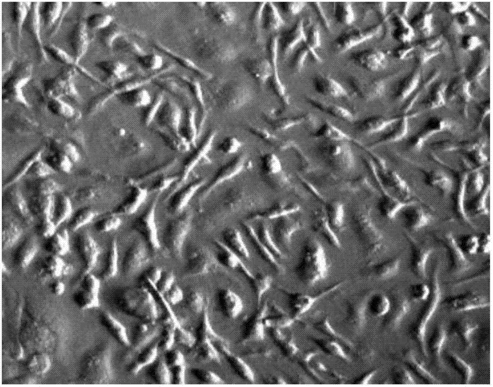
[0059] Experimental example 1: microscope observation

[0060] Obtained mouse hepatic sinusoidal endothelial cells were observed under an inverted microscope. Under a low magnification microscope, the living cells were bright, round, plump, with good transparency and clear nuclei ( figure 1 ), evaluating each mouse can obtain about 2×10 6 liver sinusoidal endothelial cells.

experiment example 2

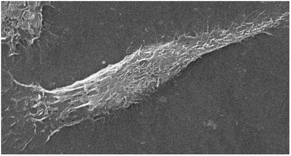
[0061] Experimental Example 2: Scanning Electron Microscope (SEM) Observation

[0062] The hepatic sinusoidal endothelial cells obtained in Example 1 were inoculated on cell slides pre-coated with rat tail collagen type I, fixed with glutaraldehyde at 4°C overnight, and dehydrated with 30%-100% alcohol gradients, with a gradient of 10%, each gradient About 2 hours, after the dehydration is completed, vacuum-dry and spray gold on the environmental scanning electron microscope ( figure 2 ), it can be seen that the cells are spindle-shaped, the nucleus is large, and the periphery is thin, and the fenestration structure of different sizes can be seen.


PUM


Abstract

The invention discloses an extraction method of mouse liver sinusoidal endothelial cells. According to the method, the separation of the liver sinusoidal endothelial cells is realized through removing kupffer cells through gadolinium chloride injection, in combination with two-step perfusion, differential centrifugation, percoll centrifugation and differential attachment. The method has the advantages of simplicity, easiness, feasibility, high cellular purity and high pick-up rate, can keep complete structure and function activity in vitro culture, and can be used for biochemistry and immunology. In addition, the method, which can use general instruments in a laboratory, is low in cost and can be popularized to most laboratories.

Description

technical field [0001] The invention relates to a method for extracting mouse liver cells, in particular to a method for extracting mouse liver sinusoidal endothelial cells. Background technique [0002] Liver sinusoidal endothelial cells (Liver Sinusoidal Endothelial Cells) are non-parenchymal cells in the liver, and play a role in the liver, such as barrier, substance metabolism, ultrafiltration endocytosis, and antigen presentation. In recent years, scholars at home and abroad have done a lot of research on the separation method of liver sinusoidal endothelial cells, but it is still very difficult to obtain liver sinusoidal endothelial cells with high purity, good cell activity and stable function, especially mouse liver sinusoidal endothelial cells. Since the mouse strains are abundant and can more fully meet the needs of various researches, it is of great significance to obtain mouse liver sinusoidal endothelial cells with good cell activity. [0003] Currently, mouse ...

Claims


Application Information

Patent Timeline
IPC IPC(8): C12N5/071
Inventor 王慧张进祥王国梁
Owner 王慧
Why Eureka
Try Eureka