In-vitro building method of cell photoaging model

A construction method and photoaging technology, applied in the fields of medicine and cell biology, can solve problems such as changing the state of cell viability, interfering with aging phenotypes, and fluctuation of cell viability, achieving the effects of good viability, good cell viability, and avoiding insufficient viability.

Inactive Publication Date: 2013-12-04
HUADONG HOSPITAL
View PDF0 Cites 5 Cited by
  • Summary
  • Abstract
  • Description
  • Claims
  • Application Information

AI Technical Summary

Problems solved by technology

The reason why this method is difficult to popularize is that first, the cell viability of cell lines is prone to fluctuate during long-term transportation, and repeated freezing and thawing will also change the state of cell viability; , which are prone to telomere shortening and will themselves develop a senescent phenotype with passage, thereby interfering with the identification of UV-induced senescent phenotypes

Method used


Image

  • In-vitro building method of cell photoaging model
  • In-vitro building method of cell photoaging model
  • In-vitro building method of cell photoaging model

Examples


Embodiment 1

[0064] Isolation and culture of MDFs

[0065] Cells are obtained from C57BL / 6 inbred mice born at 18-24 hours, and mice with ruddy complexion and normal peristalsis should be selected.

[0066] (1) Separate the back skin: After ether anesthesia, immerse the mouse in 75% ethanol for about 10 minutes, wash off the residual ethanol with DMEM, place it in a petri dish, separate the back skin with scissors, and cut off the skin tissue for about 10 minutes. 1cm×1cm, immediately placed in 2% neutral protease, digested at 37°C for 4 hours;

[0067] (2) Digestion of the skin: Take out the skin, separate the epidermis and dermis with tweezers, cut the dermis into pieces, place in 0.1% collagenase, and digest on a constant temperature shaker at 37°C for 2 hours until the tissue blocks basically disappear. Centrifuge at 1500rpm for 5 minutes to collect the precipitate;

[0068] (3) Inoculation: discard the supernatant, and make the cells with DMEM (Gibco, Gland Island, NY, USA) culture ...

Embodiment 2

[0072] UVB irradiation

[0073] (1) Preparation of UVB lamps: Wipe the UVB lamps with 75% ethanol, and pre-sterilize them in an ultra-clean bench for 30 minutes.

[0074] (2) Measurement of UVB radiation dose: Centrifuge tube racks are placed on both sides of the lamp tube to make it 30cm away from the ultra-clean table plane, and the UVB dose is measured with a Lutron UV light meter (Lutron, Taiwan) (the instrument display unit is mJ / cm 2 s), calculate the required irradiation time according to the measured data (irradiation time = 120 / UVB measured dose (calculated value in seconds));

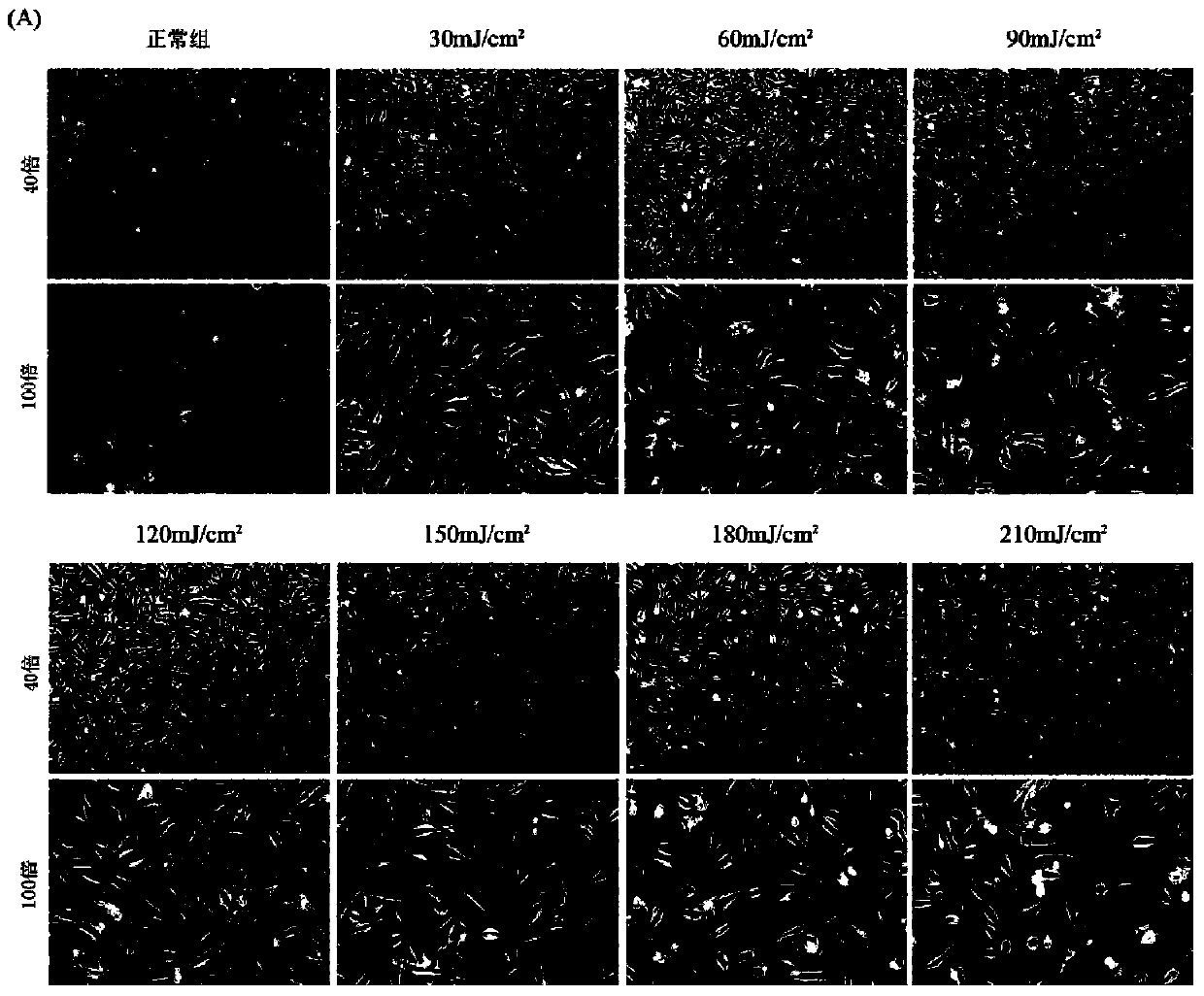
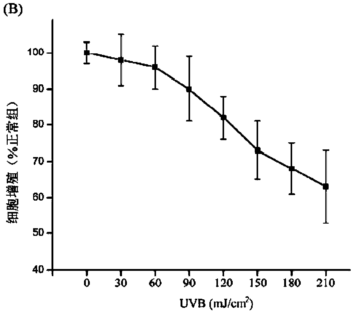
[0075] (3) UVB irradiation: when the degree of confluence of the mouse skin fibroblasts prepared in Example 1 for passage 1-3 (the figure is P2 generation) is 50%, remove the culture medium and cover with a thin layer PBS, open the cover, place it directly under the UVB lamp for the first irradiation, the irradiation dose is 120mJ / cm 2 . After the irradiation, remove the PBS, add 10ml DMEM+...


PUM


Abstract

The invention belongs to the field of medicine and cytobiology and relates to an in-vitro building method of a cell photoaging model. The method includes the following steps: (1), obtaining newborn mouse skin fibroblast primary cells, and using DMEM+10%FBS for culture in vitro; (2), when the mouse skin fibroblast primary cells grow to 80%, going down to the future generation, allowing the cells to adherently grow for 24 hours, performing first-time medium-wave ultraviolet irradiation, repeatedly irradiating for four times in each 12 hours, finally irradiating for 24-72 hours to obtain a mouse skin fibroblast cell photoaging model. Mouse-source cells are adopted, so that a lot of primary cells can be obtained only by one-time material taking; the defects of repeated cryopreseration and shortage of activity of cell lines are overcome; cell activity is good in in-vitro culture, so that no interference can be generated on a UVB-induced aging phenotype within limited generations; time needed for modeling is short, only two days are needed; effect is stable, and the aging phenotype can be observed for 24 hours after final irradiation.

Description

technical field [0001] The invention belongs to the fields of medicine and cell biology, and relates to an in vitro construction method of a cell photoaging model. Background technique [0002] Aging of the skin can be divided into two types, namely natural aging and photoaging. The former refers to the aging process controlled by the internal genes of the body as the age increases. It is only related to time and cannot be avoided by any organism. It is manifested as fine wrinkles, uniform pigmentation, loss of elasticity, loose skin and sagging. Photoaging is mainly caused by exogenous aging-promoting factors, including smoking, alcoholism, environmental pollution, and ultraviolet exposure. After long-term exposure to these environmental factors, the skin can accelerate aging and show characteristics different from endogenous aging, including coarse wrinkles, abnormal pigmentation, obvious loss of elasticity, and rough skin texture. These changes are rooted in changes in ...

Claims


Application Information

Patent Timeline
Patent Type & Authority Applications(China)
IPC IPC(8): C12N13/00C12N5/071
Inventor 刘天一曾继平毕波陈亮
Owner HUADONG HOSPITAL
Why Eureka
Try Eureka