Method for separating sperm in sperm and epithelial cell mixed stain by using immunological magnetic beads

An epithelial cell and immunomagnetic bead technology, applied in animal cells, germ cells, vertebrate cells, etc., can solve the problems of unfavorable basic laboratory popularization, low degree of automation, excessive digestion, etc., to avoid mixed typing problems, simplify The tedious process, the effect of strong capture ability

Inactive Publication Date: 2014-02-19
CHONGQING MUNICIPAL PUBLIC SECURITY BUREAU
View PDF2 Cites 13 Cited by
  • Summary
  • Abstract
  • Description
  • Claims
  • Application Information

AI Technical Summary

Problems solved by technology

[0003] In the first approach, scholars at home and abroad have established the following methods: first (Gill et al.) use the sperm outer envelope and nuclear membrane rich in protamine and thiol protein cross-linked network structure, which has stronger resistance than somatic cells Due to the structural characteristics of stress, the cells in the mixed plaque are digested in two steps, that is, the first step is to digest the somatic cells, and the second step is to digest the sperm; practice has proved that the first step of the two-step digestion method is incompletely digested or the sperm is The problem of over-digestion leads to mixed typing, female typing or no results after DNA testing; at the same time, the two-step digestion method requires two digestions, which takes a long time, requires repeated centrifugation, and has a low degree of automation; The one-step digestion method has been improved, but still can not solve the above problems; the second is (Chen et al.) taking advantage of the characteristics of the different sizes of sperm and somatic cells, the nylon filter membrane with tiny meshes can be used to filter the small volume and oval The sperm are separated from the large and flat epithelial cells, but this method also requires a strong first step of digestion, which often results in the sperm being overdigested and unable to obtain male typing; the third is (Elliot et al.) According to the sperm and body The difference in cell morphology and structure, combining optical microscopy technology with laser cell dissection technology, using laser capture microdissection technology to select and dissect sperm cells under a microscope, and obtain single male DNA typing, but this method needs to be combined with laser capture microdissection. Micro-dissection instruments and low-system amplification instruments require harsh experimental conditions, making it difficult to inspect batch samples, and it is not conducive to the popularization of grassroots laboratories

Method used


Image

  • Method for separating sperm in sperm and epithelial cell mixed stain by using immunological magnetic beads
  • Method for separating sperm in sperm and epithelial cell mixed stain by using immunological magnetic beads
  • Method for separating sperm in sperm and epithelial cell mixed stain by using immunological magnetic beads

Examples


Embodiment 1

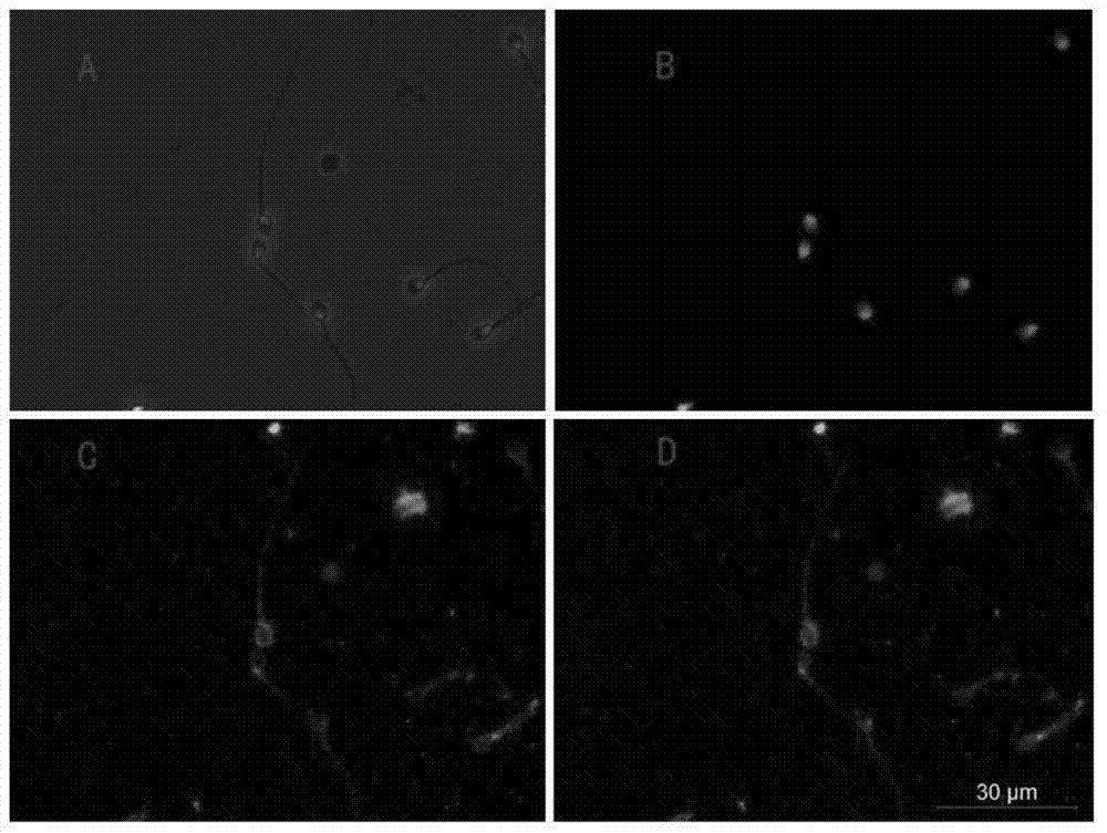
[0036] Example 1. Screening of sperm surface stable and specific membrane protein antibodies

[0037] Take 0.2 mL of sperm cell and oral epithelial cell suspension (about 10 4 pcs) on the adhesive glass slide, placed in an electric thermostat oven at 37°C to dry or air-dried at 20-25°C, fixed with fixative solution (4%-6% paraformaldehyde, 0.01-0.02mM EDTA) for 10-15 minutes , 0.5% ~ 1% Triton-100 transparent for 15 ~ 20 minutes, put it into the repair solution (0.01 ~ 0.02mol / L citrate, pH6.0, 0.01 ~ 0.02mol / L EDTA) and boil in a microwave oven for 10 ~ After 15 minutes, goat serum (Santa Cruz Biotechnology, USA) was blocked for 15 minutes, then anti-sperm membrane protein antibody (MOSPD3 or Anti-SPAG8) was added to incubate at 4°C for 12 hours, and AlExa Fluor488-labeled anti-mouse IgG (green under a fluorescent microscope) was added. Fluorescence), incubate in an electric thermostat at 37°C for 1 hour, stain with DAPI for 15 minutes, and observe under a fluorescent micros...

Embodiment 2

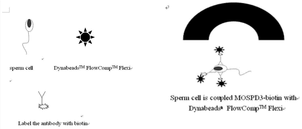
[0038]Embodiment 2, a method for immunomagnetic beads separation of sperm in mixed spots of epithelial cells, completed according to the following steps:

[0039] 1) Biotin-labeled anti-sperm membrane protein antibody:

[0040] Add 300 μL of Sulfo-NHS-LC-Biotin at a concentration of 1 mg / mL to 1 mL of MOSPD3 antibody at a concentration of 5 mg / mL or Anti-SPAG8 antibody at a concentration of 5 mg / mL, mix well in a centrifuge tube and react at 4°C for 72 hours ;Put the liquid in the centrifuge tube into the dialysis bag, add 1L of 0.01mol / L, pH7.2 PBS and dialyze at 4°C for 16 hours; get MOSPD3-biotin conjugate or Anti-SPAG8 biotin conjugate; UV / spectrophotometry The absorbance at 280nm was detected by the meter, and the measured antibody concentrations were 0.75mg / mL and 0.59mg / mL, respectively, and the molar concentration was 5.01×10 -6 mol / L, 3.921×10 -6 mol / L, the molar concentration of biotin detected by biotin quantification kit (Pierce Corporation, USA) was 1.02×10 -5 ...

Embodiment 3

[0049] Embodiment 3. A method for separating sperm from the mixed spot of epithelial cells by immunomagnetic beads is completed according to the following steps:

[0050] 1) Biotin-labeled anti-sperm membrane protein antibody:

[0051] Add 400 μL of Sulfo-NHS-LC-Biotin with a concentration of 1 mg / mL to 1 mL of MOSPD3 antibody at a concentration of 5 mg / mL, mix well in a centrifuge tube and react at 4°C for 48 hours; add the liquid in the centrifuge tube to a dialysis bag, add 1L of 0.01mol / L, pH 7.2 PBS was dialyzed at 4°C for 12 hours; the MOSPD3-biotin conjugate was obtained;

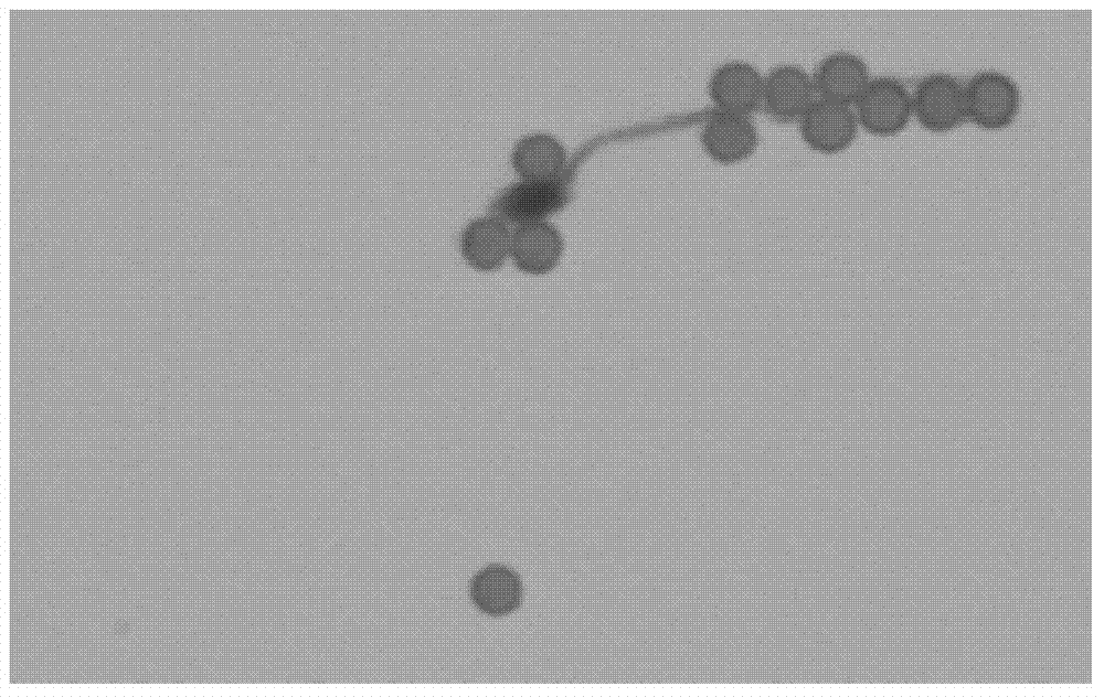
[0052] 2) Combination of sperm and antibody:

[0053] Mixed spots (containing 10 4 sperm with 10 5 Add 0.2 μL of biotin-labeled MOSPD3 antibody to the MOSPD3 antibody, place it on the Thermomixer comfort at 4°C and incubate at 800rpm for 18 hours; centrifuge at 5000rpm for 3 minutes, discard the supernatant and add 400uL 4°C pre-cooled 0.01mol / L PBS, 0.5 %BSA, 2mM EDTA, pH7.2 washing solution, af...


PUM

PropertyMeasurementUnit
diameteraaaaaaaaaa

Abstract

The invention discloses a method for separating sperm in a sperm and epithelial cell mixed stain by using immunological magnetic beads, which comprises the following steps: 1) labeling an anti-sperm membrane protein antibody with biotin; 2) combining sperm and the antibody; 3) combining magnetic beads and the antibody-sperm complex; 4) separating the sperm and an epithelial cell; and 5) separating the sperm and the magnetic beads. According to the method, the anti-sperm membrane protein antibody is labeled with the biotin, and coupling to the biotin is realized through avidin (coated on the magnetic beads), thus successfully constructing the magnetic bead-avidin-biotin-antibody-membrane antigen (sperm) complex; and a related reagent system is optimized, and a corresponding operation procedure is established, thus improving energy efficiency of mixed stain examination. The method has the advantages of fewer procedures, simple operation, convenient popularization and the like, and can provide theoretical and practical foundations for automatic examination.

Description

technical field [0001] The invention relates to a method for separating cells by immunomagnetic beads, in particular to a method for separating sperm in mixed spots of sperm and epithelial cells by immunomagnetic beads. Background technique [0002] Mixed spots containing semen are the most common biological evidence in rape cases, and their components include male cells (such as sperm, penile exfoliated cells, white blood cells in seminal plasma, etc.) and female cells (such as vaginal epithelial cells, various cells in blood etc.); how to separate and obtain the sperm of the male component in the mixed spot, that is, the suspect’s sperm, and carry out DNA typing is the key to case detection and litigation; at present, there are three ways to solve the DNA typing of the mixed spot: one is to separate the mixed spot from different Individual cells; the second is to design Y chromosome-specific DNA locus primers, and use PCR to specifically amplify the male component fragment...

Claims


Application Information

Patent Timeline
Patent Type & Authority Applications(China)
IPC IPC(8): G01N33/53
CPCC12N5/061
Inventor 李红卫王清山李学博封宇宁淑华王业全
Owner CHONGQING MUNICIPAL PUBLIC SECURITY BUREAU
Why Eureka
Try Eureka