Electrostatic spinning production method for ultrathin porous composite-nanofiber bionic Bruch's film

A composite nanofiber, electrospinning technology, applied in the field of biomedical engineering materials, to achieve the effect of promoting cell adhesion, increasing biocompatibility, and increasing cell adhesion

Active Publication Date: 2014-03-26
WENZHOU MEDICAL UNIV +1
View PDF6 Cites 7 Cited by
  • Summary
  • Abstract
  • Description
  • Claims
  • Application Information

AI Technical Summary

Problems solved by technology

[0006] In summary, there is no biomimetic Bruch's membrane prepared by nano-bionic methods, which has a porous and ultra-thin structure with staggered nanofibers and cell recognition signal sequences.

Method used


Image

  • Electrostatic spinning production method for ultrathin porous composite-nanofiber bionic Bruch's film
  • Electrostatic spinning production method for ultrathin porous composite-nanofiber bionic Bruch's film
  • Electrostatic spinning production method for ultrathin porous composite-nanofiber bionic Bruch's film

Examples


Embodiment 1

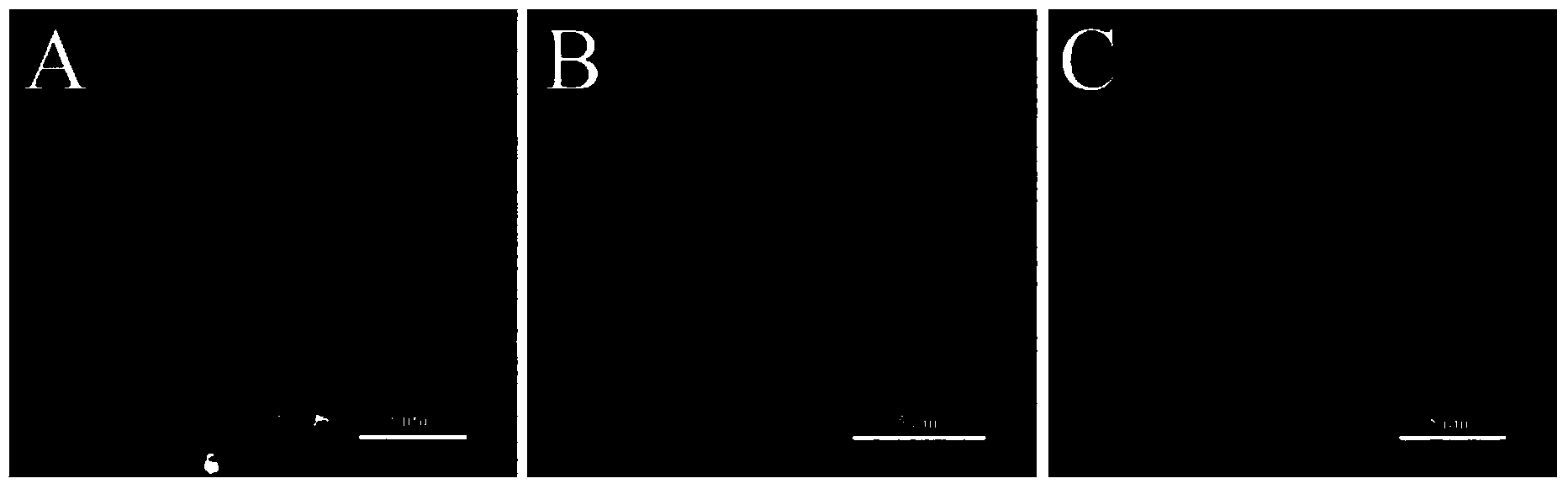
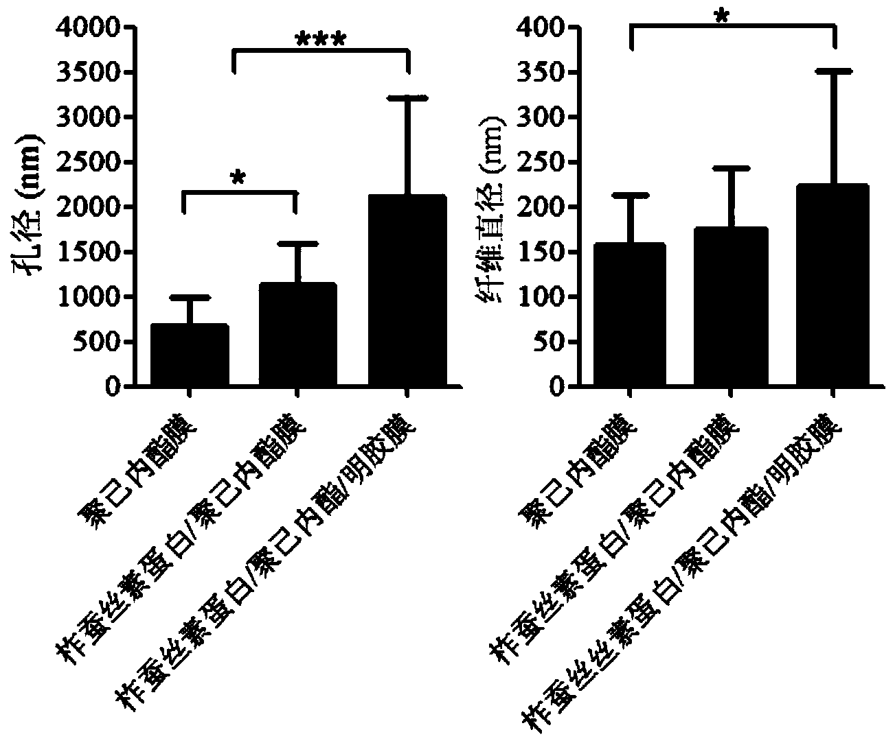
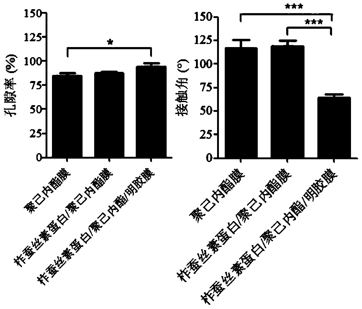
[0037] Embodiment 1 ultrathin porous polycaprolactone nanofiber biomimetic Bruch's film

[0038] The preparation method comprises the following steps:

[0039] 1. Preparation of electrospinning solution: Weigh 2.5 g of polycaprolactone, add 10 mL of 98% formic acid solution to a 50 mL Erlenmeyer flask, and stir overnight at room temperature to obtain an electrospinning solution;

[0040] 2. Electrospinning process: put the electrospinning solution into the syringe, and the syringe is equipped with a blunt needle with a diameter of 0.6mm. Electrospinning is performed under the conditions of voltage 12-20kv, collection distance 10-20cm, and extrusion speed 1-5mL / h. ;The preferred voltage is 18kv, the collection distance is 15cm, and the extrusion speed is 2mL / h;

[0041] 3. Post-processing: After the electrospinning is completed, place the electrospun membrane in a fume hood for 1-4 hours until the residual organic solvent is completely volatilized;

[0042] 4. Obtain ultra-th...

Embodiment 2

[0043] Example 2 Preparation of ultra-thin porous tussah silk fibroin / polycaprolactone composite nanofiber biomimetic Bruch's membrane

[0044] The preparation method comprises the following steps:

[0045] 1. Preparation of electrospinning solution: Weigh 0.125g of tussah silk fibroin protein and 2.125g of polycaprolactone into a 50mL conical flask, add 10mL of 98% formic acid solution, and magnetically stir overnight at room temperature to obtain an electrospinning solution;

[0046] 2. Electrospinning process: put the electrospinning solution into the syringe, and the syringe is equipped with a blunt needle with a diameter of 0.6mm. Electrospinning is performed under the conditions of voltage 12-20kv, collection distance 10-20cm, and extrusion speed 1-5mL / h. ;The preferred voltage is 18kv, the collection distance is 15cm, and the extrusion speed is 2mL / h;

[0047] 3. Post-processing: After the electrospinning is completed, place the electrospun membrane in a fume hood for ...

Embodiment 3

[0049] Example 3 Preparation of Ultrathin Porous Tussah Silk Fibroin / Polycaprolactone / Gelatin Composite Nanofiber Biomimetic Bruch's Membrane

[0050] The preparation method comprises the following steps:

[0051] 1. Preparation of electrospinning solution: Weigh 0.125g of tussah silk fibroin protein, 2.125g of polycaprolactone, and 0.25g of gelatin in a 50mL conical flask, add 10mL of 98% formic acid solution, and stir overnight at room temperature with magnetic force to obtain electrospinning silk solution;

[0052] 2. Electrospinning process: put the electrospinning solution into the syringe, and the syringe is equipped with a blunt needle with a diameter of 0.6mm. Electrospinning is performed under the conditions of voltage 12-20kv, collection distance 10-20cm, and extrusion speed 1-5mL / h. ;The preferred voltage is 18kv, the collection distance is 15cm, and the extrusion speed is 2mL / h;

[0053] 3. Post-processing: After the electrospinning is completed, place the electr...


PUM


Abstract

The invention belongs to the field of biomedical engineering materials, and discloses a production method for an ultrathin porous composite-nanofiber bionic Bruch's film. The production method includes dissolving raw materials in organic solvent to obtain electrostatic spinning solvent, and performing electrostatic spinning on the electrostatic spinning solvent to obtain a composite-nanofiber film; obtaining the ultrathin porous composite-nanofiber bionic Bruch's film after drying of the composite-nanofiber film. The ultrathin porous composite-nanofiber bionic Bruch's film includes a polycaprolactone nanofiber bionic Bruch's film, a tussah silk fibroin / polycaprolactone composite-nanofiber bionic Bruch's film or a tussah silk fibroin / polycaprolactone / gelatin composite-nanofiber bionic Bruch's film. Products produced by the method can serve as carriers for retinal pigment epithelium transplantation.

Description

technical field [0001] The invention belongs to the field of biomedical engineering materials, and in particular relates to an ultrathin and porous composite nanofiber bionic Bruch's membrane prepared by electrostatic spinning technology. Background technique [0002] Age-related macular degeneration (AMD) is a major blinding eye disease in my country, the main cause of which is the abnormal structure or function of the retinal pigment epithelium (RPE), which eventually leads to the apoptosis of retinal photoreceptor cells and leads to neurological damage. Irreversible degeneration of the retina, there is still no way to cure it. RPE transplantation is one of the most promising approaches to treat this disease. Preparation of ideal RPE cell transplantation carrier becomes the key to this technology. The natural RPE cells are located on Bruch's membrane, which is located between the neural retina and the choroid. Bruch's membrane not only provides a microenvironment for the...

Claims


Application Information

Patent Timeline
Patent Type & Authority Applications(China)
IPC IPC(8): D04H1/4382D04H1/728
Inventor 金子兵向萍李敏朱颖
Owner WENZHOU MEDICAL UNIV
Why Eureka
Try Eureka