Magnetic hydroxyl apatite/polymer tridimensional porous stent material with oriented magnetic field and preparation method thereof

A hydroxyapatite, orientation magnetic field technology, applied in medical science, prosthesis, etc., can solve the problems of no orientation magnetic field, no macroporous structure, unsatisfactory osteoinductivity, etc., to promote the formation of new bone, The effect of improving osteoinductivity and improving osseointegration performance

Inactive Publication Date: 2014-10-29
SHANGHAI NORMAL UNIVERSITY
View PDF6 Cites 15 Cited by
  • Summary
  • Abstract
  • Description
  • Claims
  • Application Information

AI Technical Summary

Problems solved by technology

However, the above-mentioned magnetic bone repair materials do not have a macroporous structure, and mainly use Fe 2 o 3 , Fe 3 o 4 Weak magnetic materials such as magnetic bone repair materials do not have an orientation magnetic field or weak magnetism, so the osteoinductivity is not ideal

Method used


Image

  • Magnetic hydroxyl apatite/polymer tridimensional porous stent material with oriented magnetic field and preparation method thereof
  • Magnetic hydroxyl apatite/polymer tridimensional porous stent material with oriented magnetic field and preparation method thereof
  • Magnetic hydroxyl apatite/polymer tridimensional porous stent material with oriented magnetic field and preparation method thereof

Examples


Embodiment 1

[0033] (1) Preparation of chitosan solution: 1 g of chitosan was dissolved in 25 mL of acetic acid solution (2 vol%), magnetically stirred for 12 h until completely dissolved, and ultrasonically removed for 1 h to remove air bubbles;

[0034] (2) Weigh 4.5g hydroxyapatite and 4.5g M-type ferrite (SrFe 12 o 19 ), placed in the chitosan solution, stirred at 450rpm for 20h until uniformly dispersed, and ultrasonically removed for 30min to remove air bubbles;

[0035] (3) Transfer the mixed solution containing chitosan, ferrite and hydroxyapatite to a 12mm×18mm (diameter×height) mold, freeze at -20°C for 10h under a uniform magnetic field of 1000Gs, and then transfer to freeze-drying freeze-dry at -80°C for 30 hours to obtain a magnetic hydroxyapatite / chitosan three-dimensional porous scaffold;

[0036] (4) At room temperature, soak the magnetic hydroxyapatite / chitosan three-dimensional porous scaffold in 100mL of 10wt% NaOH for 24 hours, wash it with deionized water for 6-8 tim...

Embodiment 2

[0040] The magnetic hydroxyapatite / chitosan three-dimensional porous scaffold obtained in Example 1 with an orientation magnetic field was tested for in vitro biological activity.

[0041] (1) Preparation of simulated body fluid: keep the temperature of deionized water in the plastic container at 37°C, add 8.035g NaCl and 0.355g NaHCO to the stirred deionized water in sequence 3 , 0.225g KCl, 0.231g K 2 HPO 4 ·3H 2 O, 0.311g MgCl 2 ·6H 2 O, 39mL1.0mol / LHCl, 0.292g CaCl 2 , 0.072g Na 2 SO 4 , then use 6.118g (CH 2 Oh) 3 CNH 2 , 1.0mol / L HCl to adjust the pH of the solution to 7.4, dilute to 1000mL, transfer to a plastic container, and store at 37°C for later use.

[0042] (2) According to the dosage ratio of 50mL / tablet, measure the simulated body fluid in a beaker, soak and transform at 37°C for 2 days, cool, wash and dry.

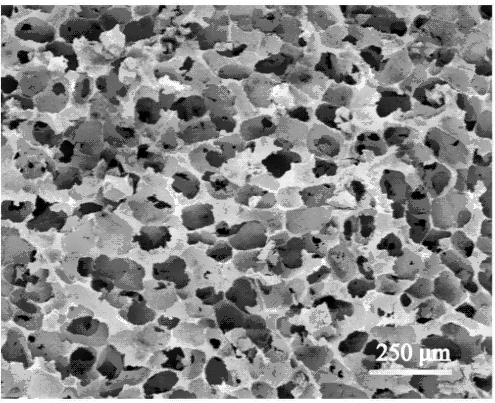
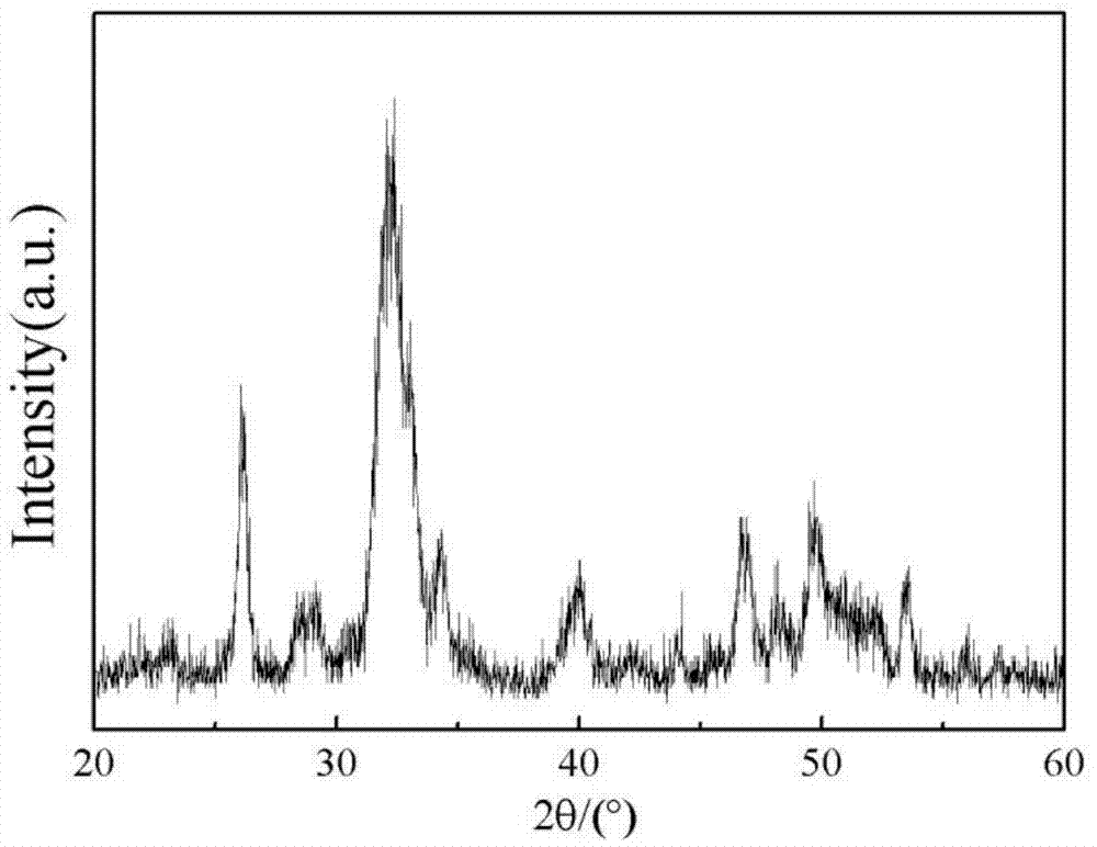
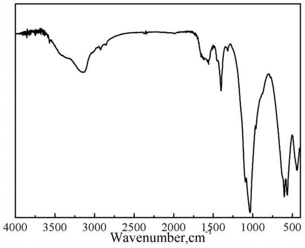
[0043] The morphology of the three-dimensional porous scaffold obtained in Example 2 was characterized, and the scanning electron microscope im...

Embodiment 3

[0045] CCK-8 method to study human bone marrow mesenchymal stem cells (hBMSCs) obtained in Example 1 with an orientation magnetic field magnetic hydroxyapatite / chitosan three-dimensional porous scaffold (Magnetic scallfolds)

[0046] And the growth of hydroxyapatite / chitosan three-dimensional porous scaffold (Normal scallfolds) material surface:

[0047] (1) Put each group of materials in a 24-well plate (the material is disc-shaped, with a diameter of 10 mm);

[0048] (2) Press 1×10 4 The density of cells / sample seeded hBMSCs on each group of materials;

[0049] (3) Add 100 μl of 5 μg / mL CCK-8 reaction solution at each time point (1d, 3d, 5d, 7d), and incubate in a constant temperature incubator at 37°C for 2 hours;

[0050] (4) Aspirate CCK-8 solution, 200 μl / well, add to 96-well plate. The OD value was measured with a microplate reader at a wavelength of 450 nm.

[0051] Figure 6 The CCK-8 method is used to evaluate the adhesion and value-added maps of human bone marr...


PUM

PropertyMeasurementUnit
pore sizeaaaaaaaaaa
porosityaaaaaaaaaa
porosityaaaaaaaaaa

Abstract

The invention relates to a magnetic hydroxyl apatite / polymer tridimensional porous stent material with an oriented magnetic field and a preparation method thereof. The stent material has an inter-communicated tridimensional porous structure, the diameter is 60 to 400 [mu]m, the porosity is 30% to 90%, and the stent material has an oriented magnetic field with an adjustable magnetic strength. The preparation method comprises the following steps: first, dissolving a bioactive polymer material into a solvent to prepare a polymer solution; then adding permanent magnetic ferrite powder and hydroxyl apatite powder into the polymer solution, evenly stirring, loading the mixed solution into a mould, freeze-drying under an externally-applied magnetic field so as to mould the solution; finally, soaking the mould into a solution, cutting, and magnetizing the cut moulds so as to obtain the tridimensional porous stent. The method is simple, the operation is convenient, the repeatability is high, and the synthesized tridimensional porous stent has the advantages of good permeability, high porosity, good biological compatibility, good osteoconduction, and good osteoconduction, and has a wide application prospect in the field of bone repairing materials.

Description

technical field [0001] The invention relates to the preparation technology of an artificial bone repair material in the field of biomedical materials, in particular to a magnetic hydroxyapatite / polymer three-dimensional porous scaffold material with an orientation magnetic field and a preparation method thereof. Background technique [0002] Large bone defects caused by trauma, osteomyelitis, bone tumors, bone cysts and other surgical resections have seriously affected the physiological functions of human bone tissue, and are currently one of the most common and difficult clinical problems in orthopedics. The current demand for suitable bone substitutes is increasing day by day. The global market of bone repair regenerative materials and related medical fields has risen from 20 billion US dollars in 2000 to 60 billion US dollars in 2011, and has increased year by year. Autologous bone is the gold standard for the treatment of bone defects. It has good histocompatibility, ost...

Claims


Application Information

Patent Timeline
Patent Type & Authority Applications(China)
IPC IPC(8): A61L27/56A61L27/50A61L27/20A61L27/24A61L27/16A61L27/18C08J9/28
Inventor 郭亚平陈伟雷勇宋真富柯勤飞
Owner SHANGHAI NORMAL UNIVERSITY
Why Eureka
Try Eureka