Preparation method and application of soluble I-type DHV (Duck Hepatitis Virus) 3D protein

A duck hepatitis virus, soluble technology, applied in the field of biochemistry, can solve the problems of difficult, laborious, and time-consuming renaturation of inclusion bodies

Active Publication Date: 2015-01-21
SICHUAN AGRI UNIV
View PDF2 Cites 5 Cited by
  • Summary
  • Abstract
  • Description
  • Claims
  • Application Information

AI Technical Summary

Problems solved by technology

Although the peptide chain of the target protein in the inclusion body is complete, it is an expressed protein in a non-natural form and has no biological activity. The dissolution of the inclusion body is very difficult, and strong denaturants such as high-concentration urea and SDS are needed to interrupt the molecule. The non-covalent bonds and ionic bonds between intramolecular and intermolecular, etc., in order to obtain active protein, protein renaturation is then required, and the renaturation of inclusion body protein after dissolution is not only time-consuming and laborious, but also has a low renaturation rate

Method used


Image

  • Preparation method and application of soluble I-type DHV (Duck Hepatitis Virus) 3D protein
  • Preparation method and application of soluble I-type DHV (Duck Hepatitis Virus) 3D protein
  • Preparation method and application of soluble I-type DHV (Duck Hepatitis Virus) 3D protein

Examples


Embodiment 1

[0025] Embodiment 1, cloning the 3D gene of type I duck hepatitis virus strain DHAV-H

[0026] Type I duck hepatitis virus strain H (DHAV-H) (see GenBank: JQ301467 for the genome sequence), E. coli DH5α strain and pET-32(a)+ vector were all preserved and provided by the Poultry Disease Control Research Center of Sichuan Agricultural University. Various molecular biology reagents were purchased from Bioreagent Company.

[0027] According to the genome sequence of DHAV-H (GenBank: JQ301467), the primers for amplifying the 3D gene were designed, and the specific primers were as follows:

[0028] P1: 5'-gg ggtacc gatcaagggaaagtagtgagcaag-3' (SEQ ID NO.1), the underline indicates the Kpn Ⅰ restriction site;

[0029] P2: 5'-acgc ctcgag tcagatcatcatgcaagctgt-3' (SEQ ID NO.2), the underline indicates the Xho I restriction site; then the designed primers were synthesized by Treasure Bioengineering (Dalian) Co., Ltd.

[0030] Take the preserved DHAV-H virus solution and make 5-fold ...

Embodiment 2

[0035] Embodiment 2, construction expresses the expression vector of type I duck hepatitis virus strain H 3D protein

[0036] The recovered DHAV-H-3D was ligated with the pJET1.2 vector, and the ligation reaction was carried out according to the instructions of the pJET1.2 cloning kit. The reaction system is shown in Table 2.

[0037] Table 2. Connection of DHAV-H-3D and pJET1.2 vector

[0038]

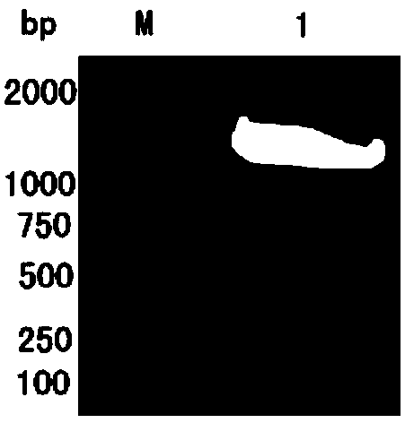
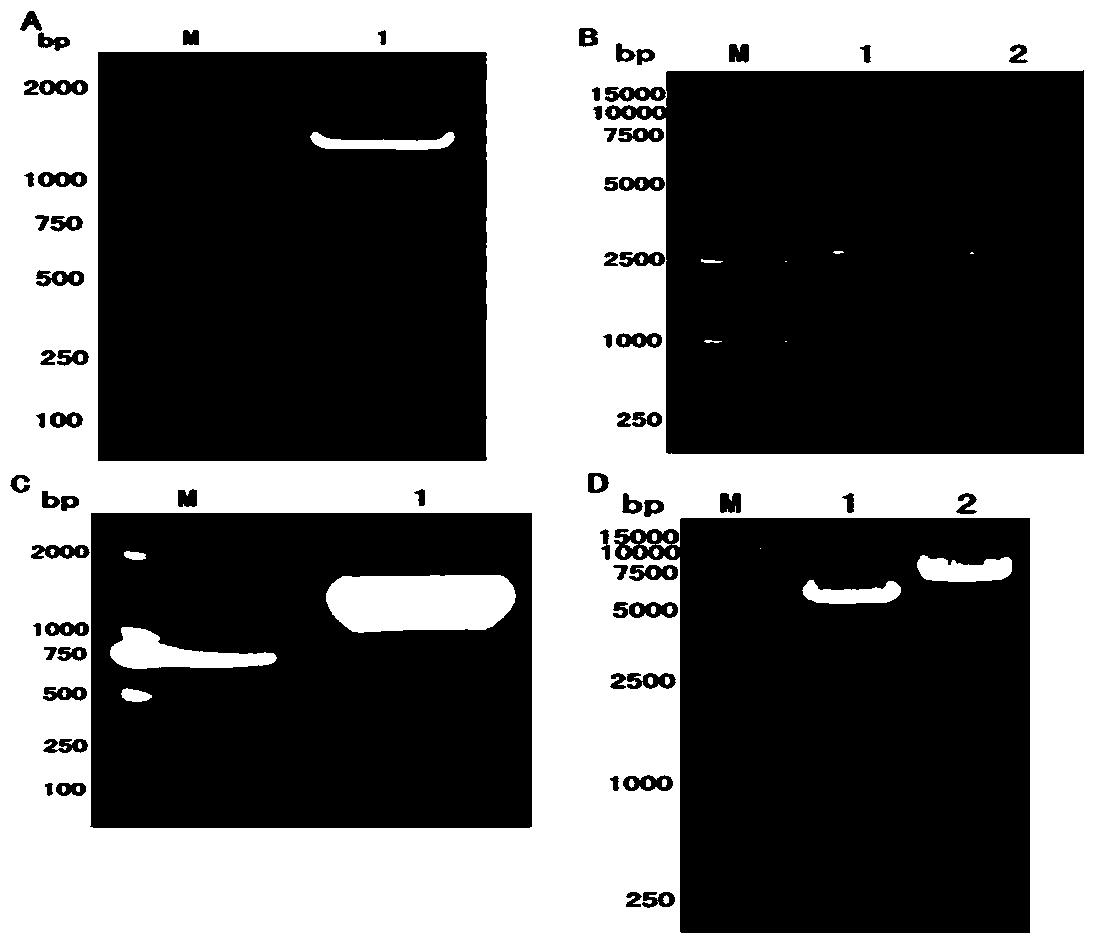
[0039] After mixing the system according to Table 2, perform transient centrifugation, and then connect at 20°C for 15 minutes. The ligation product was transformed into DH5α competent cells, and screened with Amp-resistant LB solid and liquid medium, and the screened colonies were detected by PCR using the sequences shown in SEQ ID NO.1 and SEQ ID NO.2 as primers, and the amplified products were Agarose gel electrophoresis, the results of figure 2 Shown in A. The results showed that positive clones were obtained by screening.

[0040] In order to further detect positive clone...

Embodiment 3

[0048] Embodiment 3, expression of type I duck hepatitis virus strain H soluble 3D protein

[0049] The expression host bacteria Escherichia coli (Escherichia coli) Rosetta, Escherichia coli BL21 and Escherichia coli BL21 (DE3) PLYS strains are all preserved by the Poultry Disease Control Research Center of Sichuan Agricultural University; the expression vector pET containing H 3D protein of type I duck hepatitis virus strain -32(a)+ / DHAV-H-3D was constructed from Example 2; various molecular biology reagents were purchased from Bioreagent Company.

[0050] The pET-32(a)+ / DHAV-H-3D plasmid obtained in Example 2 was transformed into BL21, Rosetta and BL21(DE3) PLYS expression bacteria respectively, and the transformed bacteria were placed in Amp-resistant LB liquid medium at 37°C and 120r Cultivate overnight under the condition of 1 / min, and expand the overnight culture and fresh Amp-resistant LB liquid medium at a volume ratio of 1:100 for about 3 hours the next day, and the O...


PUM

PropertyMeasurementUnit
diameteraaaaaaaaaa
concentrationaaaaaaaaaa

Abstract

The invention discloses a preparation method and application of soluble I-type DHV (Duck Hepatitis Virus) 3D protein. The preparation method comprises the steps as follows: firstly, the 9-1376 th nucleotide, shown in SEQ ID NO.3, is connected to the polyclone restriction enzyme cutting site of a pET-32(a)+ carrier; secondly, escherichia coli Rosetta, BL21 or BL21(DE3)PLYS is adopted as a host bacterium; thirdly, after the activation of the host bacterium in an Amp resistant LB culture medium, IPTG is put in additionally until the concentration reaches 0.2-1.0 mmol / L, and then the host bacterium is subjected to inducible expression for 4-12 hours at the temperature of 20-39 DEG C. According to the preparation method provided by the invention, the obtained I-type DHV 3D protein is soluble and has immunogenicity, can stimulate a duck body to generate an antibody for the I-type DHV 3D protein, can be used for detecting the serum sample of I-type DVH (Duck Viral Hepatitis) or a reagent containing the antibody for I-type DHV 3D protein, and has great significance for the diagnosis of the I-type DVH and the researches on vaccine for the I-type DVH.

Description

technical field [0001] The invention belongs to the field of biochemistry, and in particular relates to a preparation method of soluble type I duck hepatitis virus 3D protein, and also relates to an application of the protein. Background technique [0002] my country is the country with the largest number of ducks in the world, and the duck breeding accounts for about 70% of the world's total. The duck industry occupies an important position in my country's agricultural economy. Therefore, it is of great social and economic significance to study duck-derived microorganisms. Duck Hepatitis Virus (DHV) can cause Duck Viral Hepatitis (DVH) in ducklings. DVH is a highly fatal infectious disease characterized by acute onset, short course of disease, high morbidity and mortality. DHV is divided into three independent serotypes that are not serologically related: type I, type II, and type III. Wherein, serum type 1 duck hepatitis virus (DHV-I) is also called duck hepatitis A vir...

Claims


Application Information

Patent Timeline
Patent Type & Authority Applications(China)
IPC IPC(8): C12N15/70C07K14/10C07K16/10C07K16/06G01N33/569
Inventor 程安春汪铭书曹乾大陈孝跃
Owner SICHUAN AGRI UNIV
Why Eureka
Try Eureka