

Microfluidic chip for instantly detecting EGFR (epidermal growth factor receptor) mutation
A microfluidic chip and substrate technology, applied in the field of clinical medicine, can solve the problem that the micro-LAMP system is not fully compatible
- Summary
- Abstract
- Description
- Claims
- Application Information
AI Technical Summary
Problems solved by technology
Method used
Image

Examples
Embodiment 1
[0072] Example 1 A microfluidic chip
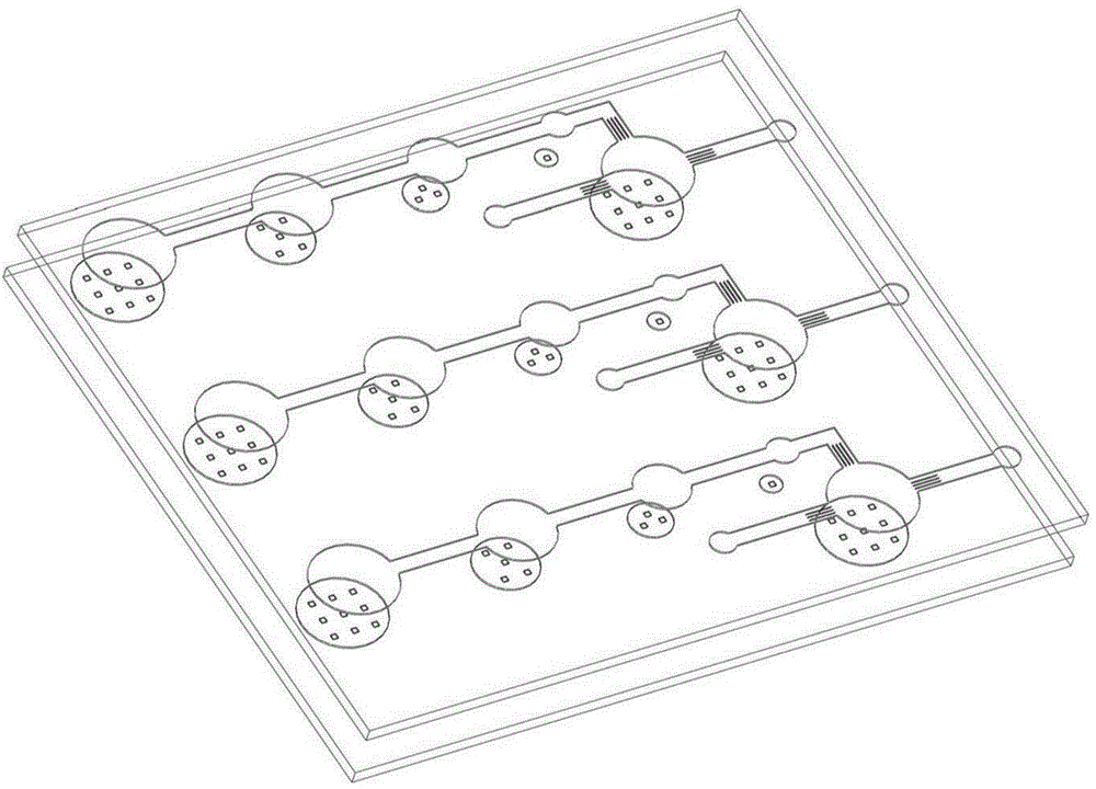
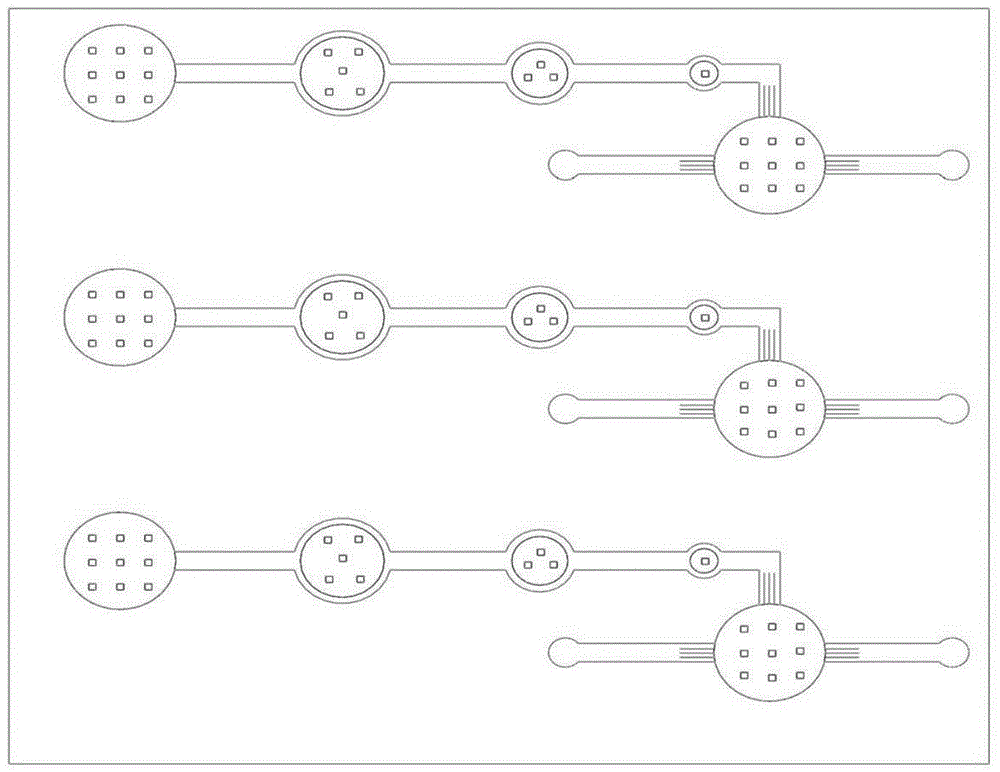
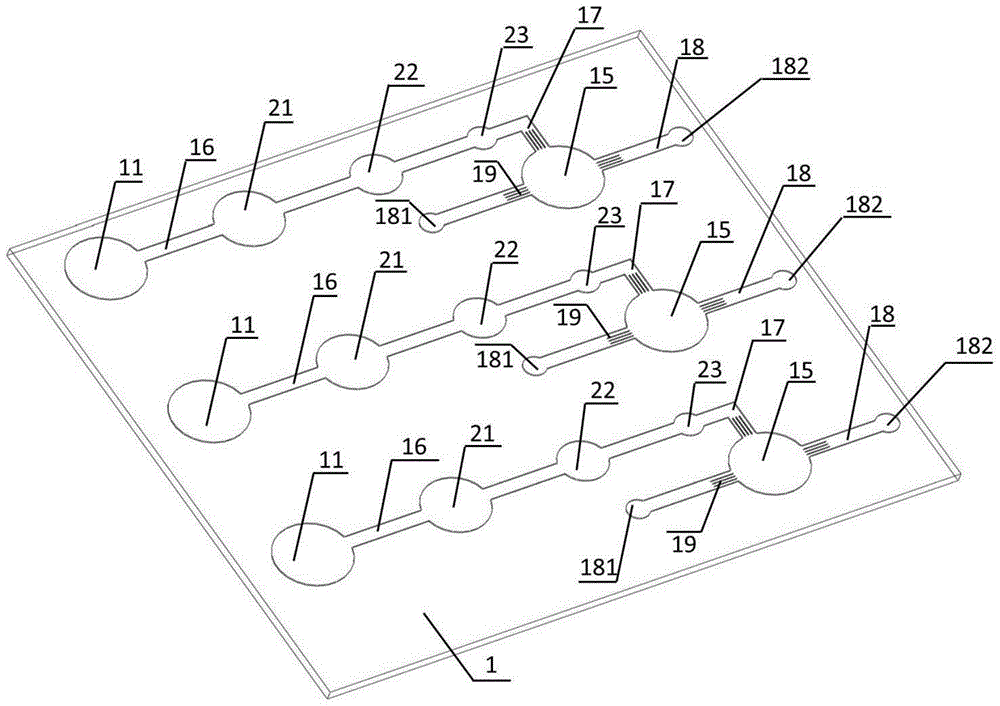
[0073] The microfluidic chip is an airtight whole formed by bonding the upper substrate 1 and the lower substrate 2; the microfluidic chip is provided with three identical and independent LAMP systems; each LAMP system includes an airbag chamber 11, A small chamber 21 equipped with constant temperature amplification buffer, a small chamber 22 equipped with primers, a small chamber 23 equipped with Bst DNA polymerase, a LAMP reaction chamber 15, a gas channel 16, a gas channel 17, a gas channel 18; the upper half of the airbag chamber 11, the upper half of the chamber 21 equipped with constant temperature amplification buffer, the upper half of the chamber 22 equipped with primers, the upper half of the chamber 23 equipped with Bst DNA polymerase, The upper part of the LAMP reaction chamber 15, the gas channel 16, the gas channel 17, and the gas channel 18 are located on the upper substrate 1, and one end of the gas channel 16 communicates...
Embodiment 2
[0096] Example 2 The preparation method of the microfluidic chip in Example 1
[0097] step:
[0098] 1. Prepare the SU-8 positive mold with microchannels and microstructures on the microfluidic chip in Example 1
[0099] Firstly, the microchannel and microstructure of the microfluidic chip were designed with computer-aided design software (Freehand); secondly, the microchannel map drawn by Freehand was printed on SU-8 glue (Microchem, model 2075) as a mask, and the The standard photolithography process is used to make the mold, and the SU-8 positive mold is obtained.
[0100] 2. PDMS replication
[0101] (1) Mix PDMS (Dow Corning, article number: 0007883528) and curing agent at a mass ratio of 10:1, pump air in a vacuum drying oven, pour it on the surface of the mold, bake at 80°C for 1 hour, and take it out;
[0102] (2) Slowly tear off the PDMS on the template after cooling, drill the injection hole and waste liquid hole at the corresponding position of the PDMS substrat...
Embodiment 3
[0107] Example 3 Research on the effect of the microfluidic chip in Example 1 in detecting EGFR gene point mutations
[0108] step:
[0109] 1. Cell collection and lysis
[0110] Take a bottle of H1975 with a confluence of 80%-90% (there is a point mutation in exon 21 of the EGFP gene on the cell genome) and a bottle of the negative control human lung adenocarcinoma cell line A549. After washing with PBS, add 1.5 mL of DNAiso reagent (Takara), shake the cell culture flask gently.
[0111] 2. Detection of point mutations in exon 21 of EGFP gene on sample DNA
[0112] (1) Use a syringe to suck out the lysate containing cell fragments, inject the above-mentioned cell lysate into the LAMP reaction chamber 15 through the sample inlet 181 in the sample LAMP system on the microfluidic chip, and let it stand for several minutes to make the nucleic acid released by cell lysis Adsorbed to the surface of the glass frit, then inject air into the sample inlet 181 with a syringe, so that...
PUM
| Property | Measurement | Unit |
|---|---|---|
| Diameter | aaaaa | aaaaa |
Abstract
Description
Claims
Application Information