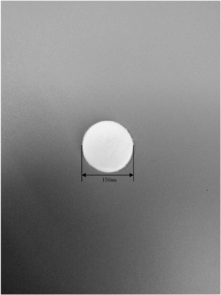
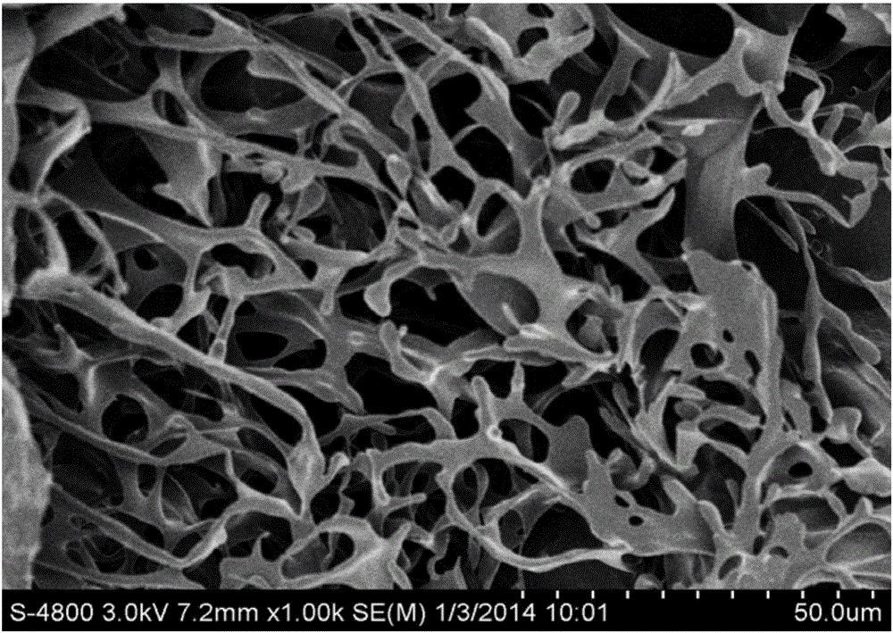
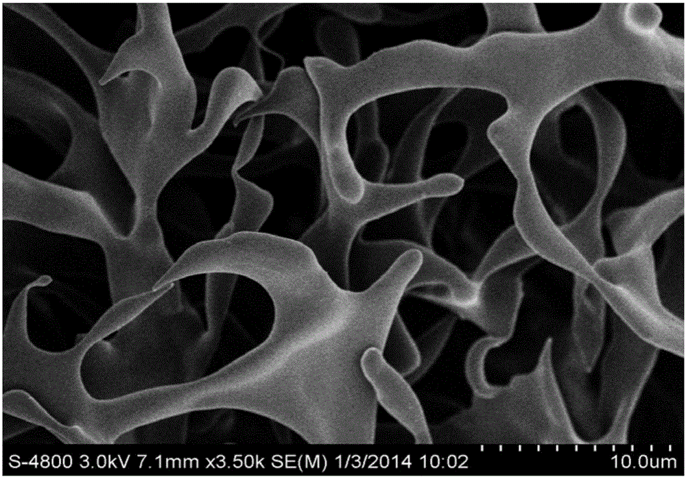
Tissue engineering cartilage stent and method for preparing same

A technology of tissue engineering and cartilage, which is applied in the field of tissue engineering cartilage scaffolds and its preparation, can solve the problems of unsatisfactory treatment effects, and achieve the effect of convenient application, good biocompatibility, and compact appearance

Active Publication Date: 2015-11-11
河北太美生物科技有限公司
View PDF4 Cites 6 Cited by
  • Summary
  • Abstract
  • Description
  • Claims
  • Application Information

AI Technical Summary

Problems solved by technology

[0004] It can be seen that there are obvious deficiencies in the methods and means of conv

Method used


Image

  • Tissue engineering cartilage stent and method for preparing same
  • Tissue engineering cartilage stent and method for preparing same
  • Tissue engineering cartilage stent and method for preparing same

Examples


Embodiment 1

[0033] The preparation method of tissue engineered cartilage scaffold of the present invention comprises the following steps:

[0034] Step 1: Extraction of type II collagen.

[0035] First, select fresh bovine cartilage, remove the periosteum with chloroform and methanol, and pulverize the fresh bovine cartilage. Next, the pulverized cartilage tissue was stirred with 0.05 mol / L Tris-HCl solution containing 4 mol / L guanidine hydrochloride, centrifuged at 4° C., and the supernatant was removed. Repeat several times to remove proteoglycan. Then, thoroughly wash the precipitate obtained by centrifugation with 0.1-0.5mol / L acetic acid, dissolve it in acetic acid of the same concentration, add pepsin to digest the precipitate for 10-18 hours, store it at 4°C and centrifuge, and centrifuge the obtained The supernatant was then digested for 5-8 hours, and the supernatant obtained above was combined to prepare a type II collagen solution. Finally, the above supernatant was salted o...

Embodiment 2

[0047] The difference between this example and Example 1 is that in Step 3, the CII-HA-CS-HAP three-dimensional cartilage primary scaffold was soaked in a mixed solution of ethanol and MES for 30 minutes at room temperature. Then, soak the primary three-dimensional cartilage scaffold in a mixed solution containing ethanol, MES, NHS, and EDC, and place it in an incubator at 25° C. for 36 hours for cross-linking. The various conditions of other experimental steps are kept unchanged, and the tissue engineering three-dimensional cartilage scaffold of the present invention is obtained.

Embodiment 3

[0049] The difference between this example and Example 1 is that in Step 3, the CII-HA-CS-HAP three-dimensional cartilage primary scaffold was soaked in a mixed solution of ethanol and MES for 30 minutes at room temperature. Then, soak the primary three-dimensional cartilage scaffold in a mixed solution containing ethanol, MES, NHS, and EDC, and put it in an incubator at 22° C. for 30 hours for cross-linking. With other conditions unchanged, the tissue engineered cartilage scaffold of the present invention is finally obtained.


PUM

PropertyMeasurementUnit
Apertureaaaaaaaaaa
Compressive strengthaaaaaaaaaa

Abstract

The invention discloses a tissue engineering cartilage stent and a method for preparing the same. The method includes a step A, extracting II-type collagen; a step B, preparing a primary CII-HA-CS-HAP three-dimensional cartilage stent by the aid of the II-type collagen, hyaluronic acid, chondroitin sulfate and hydroxyapatite; a step C, crosslinking the primary CII-HA-CS-HAP three-dimensional cartilage stent by the aid of an EDC/NHS system and freezing and drying the CII-HA-HAP three-dimensional cartilage stent to obtain the CII-HA-CS-HAP three-dimensional tissue engineering cartilage stent. The tissue engineering cartilage stent and the method have the advantages that the tissue engineering cartilage stent is excellent in biocompatibility, low in toxicity and easy to degrade, and degradation products are nontoxic; the tissue engineering cartilage stent is compact in appearance, and various performance parameters are adaptive to repairing cartilage tissues of human bodies; the constructed tissue engineering cartilage stent is made of wide material sources and easy to shape, and can be used for repairing wide-area wound, and the like.

Description

technical field [0001] The invention belongs to the technical field of tissue engineering, and in particular relates to a tissue engineering cartilage scaffold and a preparation method thereof. Background technique [0002] Articular cartilage is an essential component of joint function. It is composed of bone tissue and its surrounding perichondrium, while cartilage tissue is composed of chondrocytes, matrix and fibers. Cartilage plays an important role in joint function. However, articular cartilage damage is a very common disease. About 80% of patients undergoing arthroscopic surgery for various reasons have cartilage damage, and 20% have severe cartilage damage. Due to the lack of vascular tissue in the cartilage, and the cartilage tissue cells are wrapped in the matrix components, they cannot migrate to the damaged site to repair the defect, resulting in poor self-healing ability of the articular cartilage after injury, and regeneration cannot be achieved. If cartilag...

Claims


Application Information

Patent Timeline
IPC IPC(8): A61L27/24A61L27/20A61L27/12A61L27/50
Inventor 周晓辉冯成宝刘浩
Owner 河北太美生物科技有限公司
Why Eureka
Try Eureka