Method for separating and extracting hUC-MSC from umbilical cord outer layer amnion tissue

A technology for outer amniotic membrane and umbilical cord, applied to bone/connective tissue cells, vertebrate cells, animal cells, etc., which can solve the confusion of separation and amplification methods, complicated steps, and the difficulty of quickly extracting large quantities of high-purity hUC-MSCs And other issues

Inactive Publication Date: 2016-03-23
郭镭 +1
View PDF5 Cites 6 Cited by
  • Summary
  • Abstract
  • Description
  • Claims
  • Application Information

AI Technical Summary

Problems solved by technology

However, the separation and amplification methods of hUC-MSCs at home and abroad are confusing, and most of the ...

Method used


Image

  • Method for separating and extracting hUC-MSC from umbilical cord outer layer amnion tissue
  • Method for separating and extracting hUC-MSC from umbilical cord outer layer amnion tissue
  • Method for separating and extracting hUC-MSC from umbilical cord outer layer amnion tissue

Examples


Embodiment 1

[0092] Example 1 Screening of composition of serum-free medium for mesenchymal stem cells

[0093] (1) Content screening of serum substitutes

[0094] Test medium: 0.1 parts by volume of β-mercaptoethanol, 10ng / ml of recombinant human basic fibroblast growth factor (b-FGF, Peprotech Company), 1 part by volume of non-essential amino acid aqueous solution (11140, Gibco Company), 1, 2, 5, 8, 10, 12, 15 or 20 parts by volume of Knockout FBS serum substitute (10828-028, Gibco Company), 89 parts by volume of a-MEM.

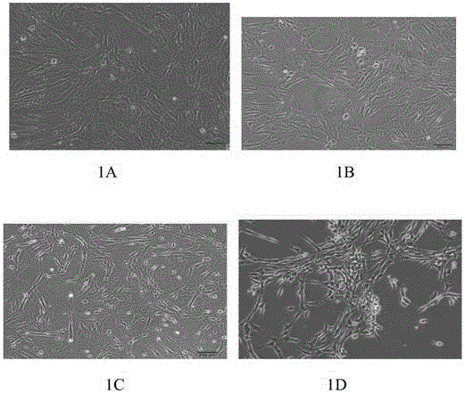
[0095] In the biosafety cabinet, the hUC-MSCs of the third generation isolated from the umbilical cord Huatong glue tissue of natural delivery newborns were collected and divided into 2×10 4 cells / cm 2 Inoculate at a density in a T75 cell culture flask, add 12-15ml of conventional commercially available medium to culture the cells. After culturing and observing that the cells have completely adhered to the wall, replace 15 mL of the test medium. Observe cell growt...

Embodiment 2

[0101] Example 2 Method for extraction of hUC-MSC by erythrocyte lysate assisted by collagenase

[0102] Collection and transportation of samples: Collect umbilical cord samples of spontaneously delivered newborns under aseptic conditions, and put them into the umbilical cord preservation and transportation solution containing penicillin sodium, streptomycin sulfate, gentamicin and amphotericin B (D-Hank's solution The concentration of penicillin sodium, streptomycin sulfate and gentamycin is 150U / ml; the concentration of amphotericin B is 300U / mL), transported to the clean cell room on ice within 6 hours;

[0103] Cleaning and disinfection of samples: In a biological safety cabinet, put fresh umbilical cord samples into a 50ml sterile centrifuge tube, wash them twice with 75% alcohol, and then wash them three times with sterile saline;

[0104] Pretreatment of umbilical cord tissue: Use ophthalmic scissors to cut the umbilical cord into small sections about 2-3cm in length,...

Embodiment 3

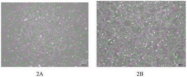
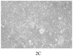
[0111] Example 3 Method for extraction of hUC-MSC by erythrocyte lysate assisted by collagenase

[0112] The red blood cell lysate used contains 10g / L NH 4 Cl and 0.1 mM Na 2 - EDTA, pH 7.2-7.4.

[0113] Carried out with reference to the method of Example 2, the digestion solution containing collagenase type IV was digested for 12 hours, the primary cells were spread on 8 culture dishes, and the mesenchymal stem cells adhered to the wall until the second day, and the confluence rate was about 7 days up to 60%, trypsinized and subcultured; after the third passage, the cell purity is greater than 99.2%, and the viability is 99.7%.


PUM


Abstract

The invention provides a method for fast separating and extracting human Umbilical Cord mesenchymal stem cells (hUC-MSC) from umbilical cord outer layer amnion tissue. The method includes the following steps that fresh collected umbilical cord tissue of a healthy newborn is taken, transported on ice in double-antibody-containing umbilical cord storing transporting liquid, washed and disinfected with 75% ethyl alcohol and normal saline, outer layer amnions are separated bluntly after blood vessels are rejected, mechanically smashed and treated with erythrocyte lysate for 3 min, IV type collagenase digestion is used, and a digestion product is cultured in a suspension mode with a serum-free culture medium after being screened through a 100-200-mesh sieve; medium change is carried out every three to five days, supernatant is taken to detect cell contamination, after the attachment rate in a plate reaches 30-70% and trypsinization is used, cell passage amplification is collected centrifugally, and confluence is carried out when the cell confluence rate reaches 90% or above, the cells are collected for cryopreservation, and the biological characteristics of hUC-MSC are detected.

Description

technical field [0001] The invention relates to an efficient and rapid separation and extraction method for umbilical cord mesenchymal stem cells, in particular to a method for extracting mesenchymal stem cells from fresh umbilical cord outer amnion tissue. Background technique [0002] Mesenchymal stem cells (mesenchymal stem cells, MSCs) are derived from the mesoderm and ectoderm in the early stages of development, have high self-renewal and multi-lineage differentiation potentials, and are widely distributed in connective tissues and interstitium of organs throughout the body. At present, studies have shown that different culture methods can cause MSCs to exhibit neuronal phenotypes, islet-like cell phenotypes, endothelial cell phenotypes, or express cardiomyocyte markers, thereby revealing the role of MSCs in immunosuppression, endocrine system regulation, and nervous system. It has broad clinical application prospects in regulating and improving cardiovascular function....

Claims


Application Information

Patent Timeline
IPC IPC(8): C12N5/0775
Inventor 郭镭
Owner 郭镭
Why Eureka
Try Eureka