A method for detecting the survival rate of cartilage tissue cells

A technology of cartilage tissue and detection method, applied in the field of biomedicine, can solve the problems of high cost and difficult realization, and achieve the effects of reliable results, avoiding damage and improving experimental efficiency.

Active Publication Date: 2019-12-24
TAISHAN MEDICAL UNIV
View PDF0 Cites 0 Cited by
  • Summary
  • Abstract
  • Description
  • Claims
  • Application Information

AI Technical Summary

Problems solved by technology

[0004] The purpose of the present invention is to provide a method for detecting the survival rate of cartilage tissue cells, which aims to solve the problems of high cost and difficult implementation in the current detection method for the repair and treatment effect of articular cartilage damage

Method used


Image

  • A method for detecting the survival rate of cartilage tissue cells
  • A method for detecting the survival rate of cartilage tissue cells

Examples


Embodiment 1

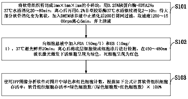
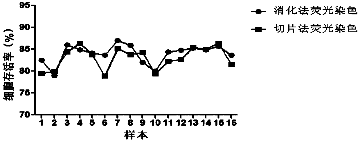
[0048] Example 1: Cut the cartilage tissue into small pieces of 1mm×1mm×1mm; digest it with 0.25% trypsin-EDTA2Na in 37℃ water bath for 80min; after centrifugation, use 0.2% type II collagenase in 37℃ water bath to continue digestion for 2h; When most of the cartilage is digested to become flocculent, add DMEM culture solution to stop the digestion and filter on a 200-mesh screen. Take the filtrate and centrifuge at 1200 rpm for 5 min. Discard the supernatant; add FDA (50mg / l) and EB (10mg) to the cell suspension. / l), incubate at 37°C for 20 minutes in the dark; after centrifugation, the bottom cells are made into cell smears and detected under 450nm wavelength laser excitation. Use IPP image analysis software to count the green and red cells in the picture to calculate the survival rate of cartilage tissue cells It is 68.37%. Tissue section fluorescence method was used to detect cell survival rate of 83.04%.

Embodiment 2

[0049] Example 2: Cut the cartilage tissue into small pieces of 1mm×1mm×1mm; digest it with 0.25% trypsin-EDTA2Na in 37℃ water bath for 40min; after centrifugation, use 0.2% type II collagenase in 37℃ water bath to continue digestion for 4h; When most of the cartilage is digested and become flocculent, add DMEM culture medium to stop the digestion and filter with a 200 mesh screen. Take the filtrate and centrifuge at 1500 rpm for 5 min. Discard the supernatant; add FDA (50mg / l) and EB (10mg) to the cell suspension. / l), incubate at 37°C for 20 minutes in the dark; after centrifugation, the bottom cells are made into cell smears and detected under 450nm wavelength laser excitation. Use IPP image analysis software to count the green and red cells in the picture to calculate the survival rate of cartilage tissue cells It is 83.97%. The cell survival rate was 83.04% by fluorescence method of tissue section.

Embodiment 3

[0050] Example 3: Cut the cartilage tissue into small pieces of 1mm×1mm×1mm; digest it with 0.25% trypsin-EDTA2Na in 37°C water bath for 20 minutes; after centrifugation, use 0.2% type II collagenase in 37°C water bath to continue digestion for 8 hours; When most of the cartilage is digested and become flocculent, add DMEM culture medium to stop the digestion and filter on a 200 mesh screen. Take the filtrate and centrifuge at 1400 rpm for 5 min. Discard the supernatant; add FDA (50mg / l) and EB (10mg) to the cell suspension. / l), incubate at 37°C in the dark for 20 minutes; after centrifugation, the bottom cells are made into cell smears and detected under 480nm wavelength laser excitation. Use IPP image analysis software to count the green and red cells in the picture to calculate the survival rate of cartilage tissue cells Is 76.59%. The cell survival rate was 83.04% by fluorescence method of tissue section.


PUM


Abstract

The invention discloses a method for detecting the survival rate of cartilage tissue cells, which comprises: cutting the cartilage tissue into small pieces, digesting in a water bath with 0.25% trypsin-EDTA2Na; centrifuging and continuing to digest in a water bath with 0.2% type II collagenase; After the cartilage is digested and becomes flocculent, add DMEM culture medium to stop the digestion, then filter through a 200-mesh screen, take the filtrate and centrifuge, and discard the supernatant; add FDA and EB to the cell suspension and incubate in the dark; use IPP image analysis software to analyze In the picture, the green and red cells are counted, and the survival rate of cartilage tissue cells is calculated according to the formula. The invention is beneficial to fully digest and separate chondrocytes, reduces the damage to chondrocytes in the process of digestion and separation, is beneficial to maintain cell activity, avoids tediousness caused by different excitation wavelengths of double fluorescent dyes, and simplifies operation; it is used for articular cartilage damage Screening of the pros and cons of treatment options and evaluation of cartilage preservation in tissue banks.

Description

Technical field [0001] The invention belongs to the technical field of biomedicine, and particularly relates to a method for detecting the survival rate of cartilage tissue cells. Background technique [0002] In recent years, with the improvement of people's living standards in our country and the full popularity of sports, the number of patients with articular cartilage injury has increased significantly. At present, there is no ideal method for repairing articular cartilage damage, and it is still an urgent medical problem to be solved internationally. At present, the clinical methods for treating articular cartilage injury include: microfracture method, autologous chondrocyte transplantation, tissue engineering cartilage transplantation, autologous and allogeneic articular cartilage transplantation, etc. The effectiveness of the above methods for the treatment of cartilage defects needs to be evaluated by reliable testing indicators, and the survival rate of cartilage tissue...

Claims


Application Information

Patent Timeline
Patent Type & Authority Patents(China)
IPC IPC(8): C12Q1/02
CPCG01N33/5005
Inventor 亓建洪宋洪强曹法民谢地胡遵杰周路
Owner TAISHAN MEDICAL UNIV
Why Eureka
Try Eureka