Device and method for endovascular optical coherence tomography - opto-acoustic - ultrasonic multimode imaging

An optical coherence tomography and blood vessel technology, which is applied in the structure, catheter, and diagnosis of ultrasonic/acoustic/infrasound diagnostic equipment. problem, to achieve the effect of simple structure, high sensitivity and good resolution

Active Publication Date: 2017-02-01
SOUTH CHINA NORMAL UNIVERSITY
View PDF7 Cites 29 Cited by
  • Summary
  • Abstract
  • Description
  • Claims
  • Application Information

AI Technical Summary

Problems solved by technology

The end of the optical fiber of this device directly emits light at 90°, the laser is divergently irradiated on the object to be measured, the laser is not converged, and there is also the problem of poor resolution of the photoacoustic image, and in the device disclosed in this technology, the coupli...

Method used


Image

  • Device and method for endovascular optical coherence tomography - opto-acoustic - ultrasonic multimode imaging
  • Device and method for endovascular optical coherence tomography - opto-acoustic - ultrasonic multimode imaging

Examples


Embodiment

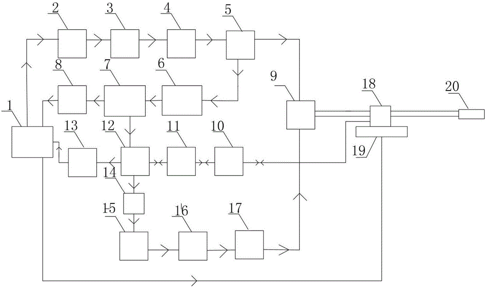
[0043] like figure 1 As shown, this embodiment discloses a device for intravascular optical coherence tomography-photoacoustic-ultrasound multimodal imaging, including a computer 1, an OCT excitation and acquisition system, a photoacoustic signal excitation and acquisition system, and an ultrasonic signal excitation With the acquisition system, and the integrated probe 20, the integrated probe is fixed on the rotation / translation platform 19 through the photoelectric slip ring 18; the optical coherence tomography OCT excitation light and the photoacoustic excitation light in the blood vessel use different light sources, and are passed through double Clad fiber for laser transmission and collection;

[0044] Wherein, the OCT excitation and acquisition system includes: FPGA board 2, superluminescent diode 3, isolator 4, first fiber coupler 5, reference arm 6, linear array CCD 7, and first acquisition card 8; The signal generated by the board excites the superluminescent diode t...


PUM

PropertyMeasurementUnit
Wavelengthaaaaaaaaaa
Bandwidthaaaaaaaaaa
Wavelengthaaaaaaaaaa

Abstract

The invention belongs to the field of blood vessel endoscopic imaging and discloses a device and method for endovascular optical coherence tomography - opto-acoustic - ultrasonic multimode imaging. The device comprises a computer, an optical coherence tomography (OCT) excitation and collection system, an opto-acoustic signal excitation and collection system, an ultrasonic signal excitation and collection system and an integrated probe. The OCT excitation and collection system comprises a field programmable logic array (PFGA) panel, a superluminescent diode, an isolator, a first optical fiber coupler, a reference arm, a linear array charge coupled device (CCD) and a first collecting card. The opto-acoustic signal excitation and collection system comprises a pulsed laser, a diaphragm, a second optical fiber coupler and a doubly clad optical fiber. The ultrasonic signal excitation and collection system comprises a pulse ultrasonic emitter/receiver, a signal amplifier, a signal filter and a second collecting card. The OCT, opto-acoustic and ultrasonic tri-modal blood vessel endoscopic imaging system integrates three imaging modes and respective advantages thereof, and multi-parameter physiological function information and multiscale structural information of a blood vessel can be obtained.

Description

technical field [0001] The invention relates to the research field of vascular endoscopy, in particular to an intravascular optical coherence tomography-photoacoustic-ultrasound multimode imaging device and method. Background technique [0002] Cardiovascular and cerebrovascular diseases are diseases with a high mortality rate, and the accuracy of blood vessel detection has higher requirements. There are modern medical methods for detecting blood vessels, including magnetic resonance (MRI) angiography, angiographic imaging (IVUS) and intravascular optical coherence tomography (OCT). The above detection methods can better present the external shape of blood vessels or internal contours, but cannot provide local details of the vessel wall and lumen. Intravascular photoacoustic imaging technology is a new type of medical imaging technology. The main principle is to irradiate tissue with pulsed laser light, and the tissue absorbs the light to generate thermoelastic waves. By im...

Claims


Application Information

Patent Timeline
IPC IPC(8): A61B5/00A61B8/12
CPCA61B5/0033A61B5/0066A61B5/0084A61B5/0095A61B8/12A61B8/44
Inventor 陈重江雷鹏杨思华邢达
Owner SOUTH CHINA NORMAL UNIVERSITY
Why Eureka
Try Eureka