Economic and practical method for extracting and separating bone tissue protein

A technology of histoprotein and separation method, which is applied in the field of molecular biology, can solve the problems of inconvenient protein extraction and analysis, high hardness and limitation of bone tissue, and achieve the effect of avoiding repeated freezing and thawing, high protein concentration and preventing degradation

Inactive Publication Date: 2017-05-31
NANJING UNIVERSITY OF TRADITIONAL CHINESE MEDICINE
View PDF4 Cites 0 Cited by
  • Summary
  • Abstract
  • Description
  • Claims
  • Application Information

AI Technical Summary

Problems solved by technology

However, in the study of bone tissue, due to the limitation of the characteristics of bone tissue itself, that is, the hardness of bone tissue is high and the content of inorganic minerals is rich, it is not convenient to extract and analyze the protein in bone tissue. In addition, the extraction of soft tissue protein in the past The method is also not applicable to bone tissue, which limits the study of related issues of bone tissue in vivo to a certain extent.
[0003] The current research on the protein level of bone tissue, especially in the research of tissue engineering bone, mainly adopts the culture of cells in bone tissue in vitro, such as osteoblasts, osteoclasts, etc., and then analyzes the protein synthesized and secreted by the cells and detection, but in fact, due to the difference in internal and external environments, the experimental results are often not completely consistent

Method used


Image

  • Economic and practical method for extracting and separating bone tissue protein

Examples


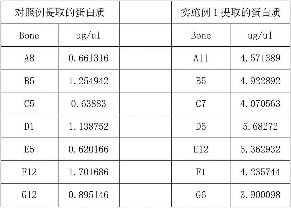
Embodiment 1

[0025] An economical and practical bone tissue protein extraction and separation method, the specific steps are as follows:

[0026] (1) Prepare the centrifuge tube and weigh it, record its mass, place the weighed centrifuge tube on ice for pre-cooling, and set aside;

[0027] (2) Take phosphatase inhibitors, protease inhibitors and phenylmethylsulfonyl fluoride and add them to the cold lysis buffer, place on ice and stir evenly to obtain the lysate. 5 μl phosphatase inhibitor, 1 μl protease inhibitor and 5 μl phenylmethylsulfonyl fluoride;

[0028] (3) Absorb physiological saline with an injection needle to wash away the bone marrow on the femur, and take 0.3 g of the femoral shaft after washing, and set aside;

[0029] (4) Place the spare femur stem in step (3) in the mortar, add liquid nitrogen to the mortar to pre-cool the mortar and the femur stem, and grind the pre-cooled femur stem for 5 minutes until the femur becomes powder shape, and continuously add liquid nitroge...

Embodiment 2

[0035] An economical and practical bone tissue protein extraction and separation method, the specific steps are as follows:

[0036] (1) Prepare the centrifuge tube and weigh it, record its mass, place the weighed centrifuge tube on ice for pre-cooling, and set aside;

[0037] (2) Take phosphatase inhibitors, protease inhibitors and phenylmethylsulfonyl fluoride and add them to the cold lysis buffer, place on ice and stir evenly to obtain the lysate. 5 μl phosphatase inhibitor, 1 μl protease inhibitor and 5 μl phenylmethylsulfonyl fluoride;

[0038] (3) Absorb physiological saline with an injection needle to wash away the bone marrow on the femur, and take 0.4 g of the femoral shaft after washing, and set aside;

[0039] (4) Place the spare femur stem in step (3) in the mortar, and add liquid nitrogen to the mortar to pre-cool the mortar and femur stem, and grind the pre-cooled femur stem for 8 minutes until the femur becomes powder shape, and continuously add liquid nitroge...

Embodiment 3

[0045] An economical and practical bone tissue protein extraction and separation method, the specific steps are as follows:

[0046] (1) Prepare the centrifuge tube and weigh it, record its mass, place the weighed centrifuge tube on ice for pre-cooling, and set aside;

[0047] (2) Take phosphatase inhibitors, protease inhibitors and phenylmethylsulfonyl fluoride and add them to the cold lysis buffer, place on ice and stir evenly to obtain the lysate. 5 μl phosphatase inhibitor, 1 μl protease inhibitor and 5 μl phenylmethylsulfonyl fluoride;

[0048] (3) Absorb physiological saline with an injection needle to wash away the bone marrow on the femur, and take 0.5 g of the femoral shaft after washing, and set aside;

[0049] (4) Place the spare femur stem in step (3) in the mortar, add liquid nitrogen to the mortar to pre-cool the mortar and femur stem, and grind the pre-cooled femur stem for 10 minutes until the femur becomes powder shape, and continuously add liquid nitrogen to ...


PUM


Abstract

The invention discloses an economic and practical method for extracting and separating bone tissue protein and belongs to the technical field of molecular biology. The method concretely comprises the following steps: placing a weighted centrifugal tube onto ice and pre-cooling; taking a phosphatase inhibitor, a protease inhibitor and phenylmethylsulfonyl fluoride, adding the three into a splitting buffer solution, and then placing the mixture onto the ice and uniformly stirring; weighting 0.3-0.5g of femoral shaft; putting the femoral shaft into a grinding kettle filled with liquid nitrogen and grinding the femoral shaft into powder; transferring the femoral shaft to a spare centrifugal tube and weighting, thereby obtaining the weight of the femoral shaft; multiplying the mass by 1 / 3, and then adding the lysate into the centrifugal tube in concentration of 0.2-0.4g / mL; shaking the mixed solution under a low temperature and then standing; placing the turbid liquid under the conditions of 4 DEG C and 3000-5000rpm and centrifuging; and absorbing the supernate to a new centrifugal tube and then split-charging and storing. According to the invention, the extraction and separation process is simple and the separated protein purity is high, so that the extraction and separation process is suitable for popularization and application in the field of biology.

Description

technical field [0001] The invention relates to the technical field of molecular biology, in particular to an economical and practical bone tissue protein extraction and separation method. Background technique [0002] At present, many problems, such as fracture healing, mechanism of bone tumor occurrence, mechanism of bone defect repair with tissue engineering bone, etc., need to be solved urgently. In the past, immunohistochemical methods were mainly used for pathophysiological research of bone tissue in vivo. With the continuous deepening of research, the explanation of related issues should gradually go deep into the protein level and gene level, which requires the detection of protein expression in bone tissue, so as to explain the occurrence mechanism of related phenomena in a more in-depth manner, so as to improve relevant theories to guide clinical practice. However, in the study of bone tissue, due to the limitation of the characteristics of bone tissue itself, tha...

Claims


Application Information

Patent Timeline
Patent Type & Authority Applications(China)
IPC IPC(8): C07K14/47C07K1/14
CPCC07K14/47
Inventor 闵文黄桂成方彭华袁翰黄正泉朱燕韩龙王磊周龙云
Owner NANJING UNIVERSITY OF TRADITIONAL CHINESE MEDICINE
Why Eureka
Try Eureka