Separation method and application of placenta-derived stem cells
A technology derived from placenta and stem cells, applied in animal cells, vertebrate cells, bone/connective tissue cells, etc., can solve problems such as poor therapeutic effect and peripheral blood vessel occlusion
- Summary
- Abstract
- Description
- Claims
- Application Information
AI Technical Summary
Problems solved by technology
Method used
Image
Examples
Embodiment 1
[0052] Example 1: Isolation and culture of placental mesenchymal stem cells
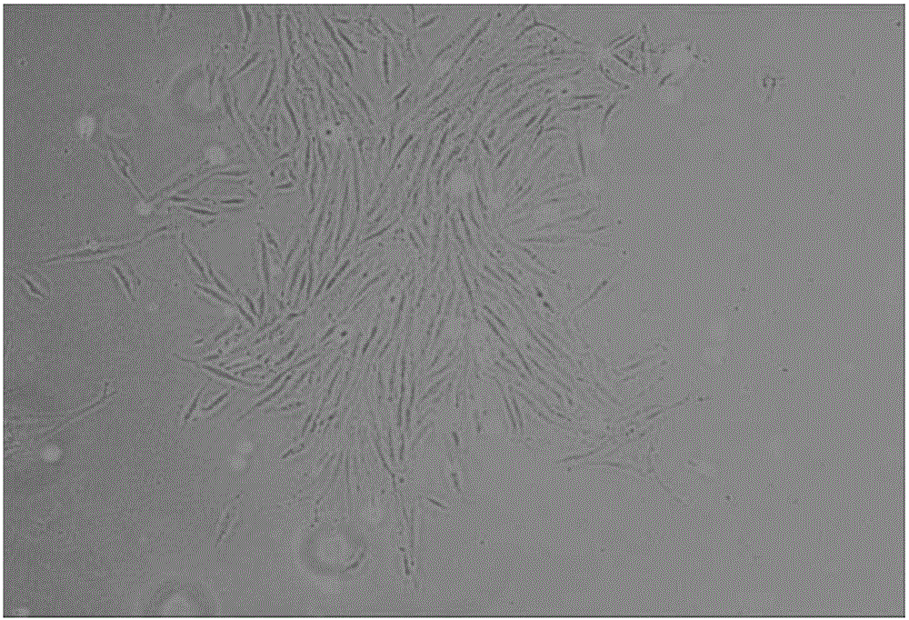
[0053] Using a new compound enzyme culture system, including 0.05mg / mL trypsin, 1mg / mL collagenase and 1mg / mL dispase (neutral enzyme), the digestive enzyme combination solution was pumped into the placenta through the umbilical vein and arteries at a speed of 50mL / hour to achieve the effect of digesting and separating cells. Digest at 37°C for 2 hours. Flushed cells were collected, washed and cultured. The culture conditions are DMEM containing 15% FBS to separate and culture placental mesenchymal stem cells PSCs are fibroblast-like, and the cells are in good condition ( figure 1 ).
[0054] 1. Reagent preparation
[0055] a) Dispase (neutral protease Ⅰ) stock solution (5mg / mL)
[0056] Dissolve the dispase in ultrapure water. The freeze-dried enzyme can be stored at 2-8 degrees until the shelf life. The aqueous solution is stored at -15 to -25 degrees. When opening the lid, be careful not to...
Embodiment 2
[0095] Example 2: Ad-HGF modified PSCs promote HGF expression
[0096] PSCs were infected with Ad-HGF at a multiplicity of infection (MOI). After 48 hours of infection, the cells were collected, and then the expression level of HGF was determined by QPCR. Moreover, after the angiography, the ischemic hindlimb muscle tissue of the rabbit was taken, and then the expressions of VEGF, bFGF and HGF in the limb muscle tissue were determined by QPCR.
[0097] Materials: DMEM medium and Ad-HGF were produced and prepared by our laboratory, and VEGF, bFGF, and HGF primers were synthesized by Aoke Company. QPCR instrument 7500 (ABI company, the United States), Green Real-time PCR Master Mix (TAKARA company, Japan).
[0098] Method and steps
[0099] 1). Extraction of tissue RNA: After the imaging, take the rabbit ischemic hindlimb muscle tissue, add liquid nitrogen to freeze and grind, add TRIZOL to lyse and extract tissue total RNA, the steps are as follows:
[0100] ① Cut out 0....
Embodiment 3
[0132] Example 3: HGF Gene Modified PSC Transplantation Promotes Angiogenesis
[0133] 3.1 Production of animal models and stem cell transplantation:
[0134] Materials: 10 New Zealand white rabbits, ordered by the Experimental Animal Center of the Academy of Military Medical Sciences (animal license number: SCXK (Beijing) 2015-0005), weighing 2-3 kg, male and female, 6 weeks old.
[0135] Method and steps
[0136] 3.1.1 Rabbits were given general anesthesia by injecting 3% pentobarbital sodium (50mg / kg) through the ear vein, and the left and right lower limbs of the experimental rabbits were used to make models.
[0137] 3.1.2 After depilation and disinfection of the left and right hind limbs, a long incision was made from the midpoint of the inguinal ligament to the knee, and a longitudinal incision was made from the inguinal ligament on both sides to the knee joint to separate the femoral artery and its branches. The femoral artery and its branches were cut off, and 1 m...
PUM
| Property | Measurement | Unit |
|---|---|---|
| weight | aaaaa | aaaaa |
| thickness | aaaaa | aaaaa |
Abstract
Description
Claims
Application Information
 - R&D
- Intellectual Property
- Life Sciences
- Materials
- Tech Scout
- Unparalleled Data Quality
- Higher Quality Content
- 60% Fewer Hallucinations
Browse by: Latest US Patents, China's latest patents, Technical Efficacy Thesaurus, Application Domain, Technology Topic, Popular Technical Reports.
© 2025 PatSnap. All rights reserved.Legal|Privacy policy|Modern Slavery Act Transparency Statement|Sitemap|About US| Contact US: help@patsnap.com


