
Spontaneous fluorescence and magnetic resonance bimodal molecular fusion small animal imaging system and method

A magnetic resonance imaging and autofluorescence technology, which is applied in the field of biomedical molecular imaging, can solve the problems of unfavorable reduction of optical tomography reconstruction problems, morbidity, difficult image resolution to meet biological experimental application research, poor magnetic compatibility of optical systems, etc. Achieve clear anatomical structure, rich information and accurate registration

Inactive Publication Date: 2017-08-22
PEKING UNION MEDICAL COLLEGE HOSPITAL CHINESE ACAD OF MEDICAL SCI
View PDF5 Cites 1 Cited by
  • Summary
  • Abstract
  • Description
  • Claims
  • Application Information

AI Technical Summary

Problems solved by technology

[0007] First, the magnetic compatibility of the optical system is poor;
[0008] Second, the magnetic resonance system has a low field strength, and the image resolution is difficult to meet the application research of subsequent biological experiments, and the magnetic resonance equipment needs to be modified;
[0009] Third, using optical fibers to detect optical signals on the surface of living organisms, the amount of measurement data is proportional to the number of fibers, so the amount of measurement data is limited, which is not conducive to reducing the pathological nature of optical tomography reconstruction problems

Method used


Image

  • Spontaneous fluorescence and magnetic resonance bimodal molecular fusion small animal imaging system and method
  • Spontaneous fluorescence and magnetic resonance bimodal molecular fusion small animal imaging system and method
  • Spontaneous fluorescence and magnetic resonance bimodal molecular fusion small animal imaging system and method

Examples


Embodiment Construction

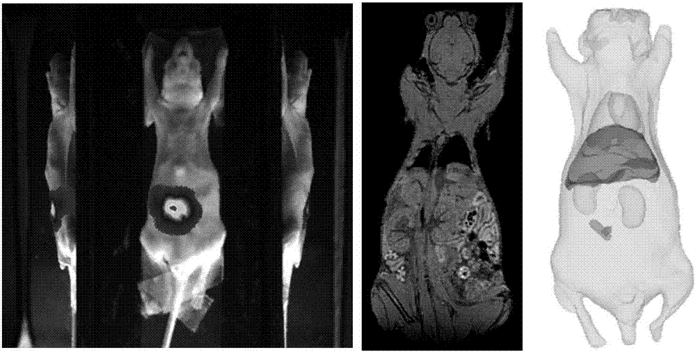
[0064] The present invention will be described in further detail below in conjunction with the accompanying drawings.

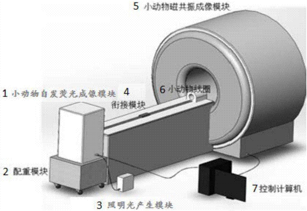
[0065] Such as figure 1 , 2 As shown, the small animal autofluorescence and magnetic resonance dual-mode molecular fusion imaging system of the present invention includes:

[0066] Small animal magnetic resonance imaging module 5, which includes a magnetic resonance examination bed;

[0067] The small animal autofluorescence imaging module 1 is placed at the end of the magnetic resonance examination table, and the small animal autofluorescence imaging module 1 uses a CCD to detect optical signals;

[0068] The connecting module 4 is placed on the magnetic resonance examination bed, and can move back and forth between the small animal autofluorescence imaging module 1 and the small animal magnetic resonance imaging module 5, and is used to realize the small animal between the autofluorescence imaging module and the magnetic resonance imaging module One-stop...


PUM


Abstract

The invention relates to a spontaneous fluorescence and magnetic resonance bimodal molecular fusion small animal imaging system and a spontaneous fluorescence and magnetic resonance bimodal molecular fusion small animal imaging method. The spontaneous fluorescence and magnetic resonance bimodal molecular fusion small animal imaging system comprises a magnetic resonance small animal imaging module, a spontaneous fluorescence small animal imaging module, a jointing module and a system control module, wherein the magnetic resonance small animal imaging module comprises a magnetic resonance examination bed; the spontaneous fluorescence small animal imaging module is placed at the tail end of the magnetic resonance examination bed; the spontaneous fluorescence small animal imaging module detects optical signals through a CCD; the jointing module achieves one-stop information acquisition of a small animal between the spontaneous fluorescence imaging module and the magnetic resonance imaging module; the system control module is used for controlling the spontaneous fluorescence small animal imaging module, the magnetic resonance small animal imaging module and the jointing module and processing the acquired magnetic resonance and fluorescence data. Through the system and the method,spontaneous fluorescence and high-field magnetic resonance bimodal imaging is adopted to provide effective molecular imaging methods for detecting abnormality in cellular and molecular levels during lesion, exploring occurrence, development and metastasis of a tumor, monitoring a therapeutic process, evaluating the therapeutic effect of a medicine and the like.

Description

technical field [0001] The invention relates to the technical field of biomedical molecular imaging, in particular to a small animal autofluorescence and magnetic resonance dual-mode molecular fusion imaging system and method. Background technique [0002] Optical and magnetic dual-mode molecular fusion imaging is one of the cutting-edge molecular imaging technologies. After nearly ten years of development, molecular imaging technology has been widely recognized and valued for its application value and potential in the field of biomedicine, and it is generally recognized that this technology is one of the important links from basic to clinical transformation. [0003] At present, emerging molecular imaging technologies are emerging in an endless stream in the world. MR (magnetic resonance, magnetic resonance), as an increasingly widely used clinical imaging device, has incomparable advantages in reflecting the body's anatomical shape and physiological function information. ...

Claims


Application Information

Patent Timeline
IPC IPC(8): A61B5/055A61B5/00G06F17/16G06F17/50G06T5/50G06T7/00
CPCA61B5/0035A61B5/0071A61B5/055G06F17/16G06F30/23G06T5/50G06T2207/10088G06T2207/20221
Inventor 金征宇薛华丹雷晶梁继民曹旭陈多芳张军蒙茗田捷
Owner PEKING UNION MEDICAL COLLEGE HOSPITAL CHINESE ACAD OF MEDICAL SCI
Learn More