Thrombin detection method based on graphene quantum dot/ erbium ion fluorescent nano probe

A fluorescent nanoprobe, graphene quantum dot technology, applied in the field of optical sensing, can solve complex price, time-consuming, expensive and other problems, and achieve the effect of good selectivity and high sensitivity

Inactive Publication Date: 2017-08-29
NANCHANG UNIV
View PDF2 Cites 3 Cited by
  • Summary
  • Abstract
  • Description
  • Claims
  • Application Information

AI Technical Summary

Problems solved by technology

However, most of the above aptamer-based thrombin detection methods require chemical modification o

Method used


Image

  • Thrombin detection method based on graphene quantum dot/ erbium ion fluorescent nano probe
  • Thrombin detection method based on graphene quantum dot/ erbium ion fluorescent nano probe
  • Thrombin detection method based on graphene quantum dot/ erbium ion fluorescent nano probe

Examples


Embodiment 1

[0019] Preparation of GQDs

[0020] (1) Add 1.0 g of carbon powder, 60 mL of concentrated nitric acid and 180 mL of concentrated sulfuric acid into a 250 mL reaction flask in sequence, and sonicate for 2 h at 100% power;

[0021] (2) The above solution was refluxed at 120 °C for 24 h, and the mixture was diluted to 800 mL with ultrapure water to obtain a dark brown solution, which was washed with Na 2 CO 3 The pH of the neutralization solution is about 7, and most of the sodium salt is removed by concentration and filtration, and the filtrate is dialyzed in 1000Da dialysis bags for 3 days in batches. The products in the dialysis bags are GQDs, and the GQDs are dispersed in ultrapure water for later use.

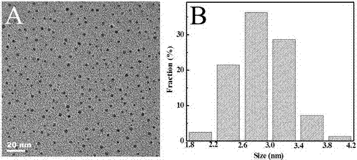
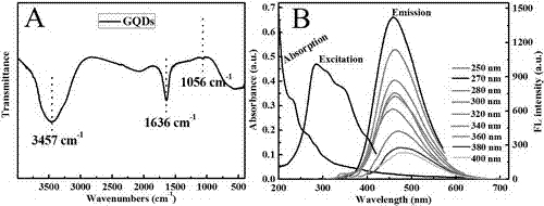
[0022] The prepared GQDs were characterized by transmission electron microscopy (TEM), infrared spectroscopy, ultraviolet-visible absorption and fluorescence spectroscopy. Depend on figure 1 A shows that the GQDs synthesized by the method of the present invention are in th...

Embodiment 2

[0024] GQDs and Er 3+ and the interaction mechanism of thrombin

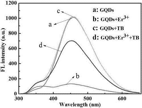
[0025] Prepare GQDs, GQDs / Er with Tris-HCl buffer solution 3+ , GQDs+TB and GQDs / Er 3+ +TB solution, add ultrapure water to bring the total volume of the test solution to 200 μL, shake it well, measure the fluorescence spectrum of the solution when the excitation wavelength is 270 nm, the final concentration of Tris-HCl buffer solution is 50 mM and the pH is 7.0, GQDs 12.5 μg / mL, Er 3+ 15 μM for TB and 20 nM for TB. Depend on image 3 It can be seen that GQDs have a strong emission peak at 456 nm (curve a); when adding Er to GQDs 3+ After that, the fluorescence of GQDs is greatly reduced (curve b); when GQDs and Er 3+ When TB was added to the mixed solution of GQDs, the fluorescence of GQDs was enhanced (curve d); however, when TB was added to the GQDs solution, the fluorescence of GQDs remained unchanged (curve c).

[0026] GQDs and Er 3+ To characterize the interaction between ( Figure 4 A), the dynami...

Embodiment 3

[0028] GQDs / Er 3+ Composite fluorescent nanoprobes for the detection of TB

[0029] 10 μL of 0.25 mg / mL GQDs solution, 20 μL of 150 μM Er 3+ solution, 20 μL of 50 mM Tris-HCl buffer solution with pH 7.4 and different concentrations of thrombin solutions were mixed, and ultrapure water was added to bring the total volume of the test solution to 200 μL, shaken and shaken, and the fluorescence of the solution was measured when the excitation wavelength was 270 nm spectrum. Depend on Figure 5 It can be seen that with the increase of TB concentration, the fluorescence of GQDs gradually increases, and the fluorescence intensity of GQDs has a good linear relationship with the TB concentration in the range of 0.1-6 nM, and the linear correlation coefficient R 2 =0.993, the detection limit of TB is 0.049 nM, which can realize the sensitive detection of TB.


PUM


Abstract

The present invention discloses a thrombin detection method based on a graphene quantum dot/ erbium ion fluorescent nano probe. Rare earth ions Er<3+> are combined with carboxylic acid groups on the surface of GQDs (graphene quantum dots) for assembly onto the surface of the GQDs to form a GQDs / Er<3+> complex to cause fluorescence quenching of the GQDs, when thrombin isadded, the GQDs / Er<3+> complex is decomposed to recover fluorescence of the GQDs, the fluorescence recovery degree of the GQDs is positively-proportional to the concentration of the thrombin, and the thrombin can be detected. The detection of the sensitivity and selectivity of the thrombin can be achieved only by fluorescence switch converting of the GQDs without functional modification of the surface of the GQDs and use of a thrombin aptamer.

Description

technical field [0001] The invention discloses a graphene quantum dot / erbium ion (GQDs / Er 3+ ) The invention relates to a thrombin detection method of a fluorescent nanoprobe, which belongs to the technical field of optical sensing. Background technique [0002] Graphene quantum dots (GQDs) refer to graphene nanosheets with a particle size of less than 100 nm, which have low toxicity, good biocompatibility, and photostability, and are widely used in substance detection, bioimaging, and fluorescence sensing. Shi et al. used GQDs-PEG-aptamer as a donor and molybdenum disulfide nanosheets as a quencher to establish a method for detecting epithelial cell adhesion molecule proteins based on GQDs fluorescent switches. Qiu et al established a Zr-based 4+ A protein kinase activity assay for multiple sequestration with phosphorylated polypeptide-GQDs complexes. Qian et al. used functionalized GQDs surface-coupled aptamers as probes to detect thrombin. However, the above GQDs-base...

Claims


Application Information

Patent Timeline
IPC IPC(8): G01N21/64
CPCG01N21/6428G01N21/6486G01N2021/6432
Inventor 梁汝萍韦甜甜邱建丁
Owner NANCHANG UNIV
Why Eureka
Try Eureka