Fluorescent cell sensor for screening inflammasome NLRP3 activators and inhibitors

An activator and inhibitor technology, applied in the field of drug screening, can solve the problems of small processing volume, unsuitability, and inability to use NLRP3 intervention agent screening, etc., to achieve the effect of improving efficiency

Active Publication Date: 2017-09-22
JIANGNAN UNIV
View PDF1 Cites 2 Cited by
  • Summary
  • Abstract
  • Description
  • Claims
  • Application Information

AI Technical Summary

Problems solved by technology

Drug screening targeting the inflammasome is the current development direction of anti-inflammatory drug research and development. However, when studying the activation and inhibition of inflammasome 3, the methods used are mainly Western, RT-PCR, etc. , these methods are cumbersome to operate and require the operator to have a certain professional quality. It usually takes two days to perform a Western experiment. Reagent consumables such as antibodies are expensive, and the amount of processing in the same batch is small, and only single-digit samples can be operated at most. Obviously such a method is not suitable and cannot be used for the screening of NLRP3 intervention agents

Method used


Image

  • Fluorescent cell sensor for screening inflammasome NLRP3 activators and inhibitors
  • Fluorescent cell sensor for screening inflammasome NLRP3 activators and inhibitors
  • Fluorescent cell sensor for screening inflammasome NLRP3 activators and inhibitors

Examples


Embodiment 1

[0043] Example 1 Construction of NLRP3-GRE vector plasmid

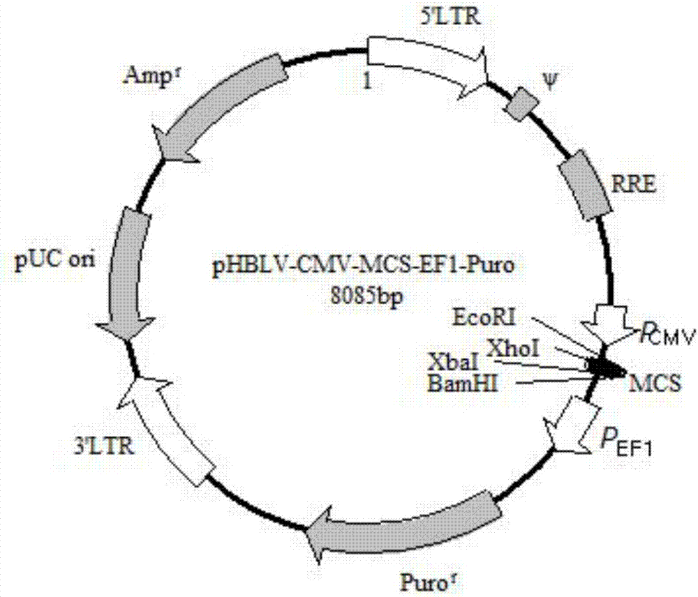
[0044] 1. The purpose is to connect the NLRP3 core promoter region with zsGREEN, and then construct pHBLV-CMVIE-EF1-Puro with ClaI / EcoRI double enzyme digestion. The vector map is as follows figure 1 shown. Since two separate sequences are spliced ​​together to form a sequence, it is necessary to design primers in segments, and then perform a full-length PCR reaction using the PCR-derived fragments as templates. The primers were designed as follows:

[0045] NLRP3-claI-EcoRI-F: gcagagatccagtttatcgatATGCTGGGGAAGTGTGTCT

[0046] zsGREE(NNLRP3)-r:ccgtgcttggactgggccaTggtggcATGGAGGGAAAAATATGCAA

[0047] zsGREE(NNLRP3)-f:CCCTCCATgccaccAtggcccagtccaagcacgg

[0048] NLRP3-claI-EcoRI-R:agaactagtctcgaggaattcttagggcaaggcggagc

[0049] 2. The pHBLV-CMVIE-EF1-Puro vector was digested with EcoRI and ClaI. The enzyme digestion system is as follows:

[0050] 40ul enzyme digestion system (2ul 400ng / ul vector, 1ul EcoRI enzyme, 1...

Embodiment 2

[0063] Example 2 Packaging of Lentivirus Containing Plasmid NLRP3-GRE

[0064] 1. Carry out massive amplification of the vector plasmid in E. coli DH5α competent cells

[0065] (1) Take 100 μl of competent cell suspension from the -70°C refrigerator, thaw it at room temperature, and put it on ice immediately after thawing.

[0066] (2) Add 5 μg of plasmid DNA solution, shake gently, and place on ice for 30 minutes.

[0067] (3) Heat shock in a water bath at 42°C for 90 seconds. Do not move the centrifuge tube during the heat shock process. After the heat shock, quickly place it on ice to cool for 3-5 minutes.

[0068] (4) Add 1mL LB liquid culture medium (without antibiotics) to the tube, pipette and mix well, and incubate at 37°C, 220rpm shaker for 1 hour, so that the bacteria can return to normal growth state and express the antibiotic resistance encoded by the plasmid Gene.

[0069] (5) Shake the above-mentioned bacterial solution, centrifuge, remove 900 μL of supernatan...

Embodiment 3

[0079] Example 3 Construction of Stably Transduced Cell Lines

[0080] 1. Infection of Thp-1 cells with lentivirus

[0081] Thp-1 cells were plated with antibiotic-free 1640+2% fetal bovine serum FBS at a cell density of 2.5×10 5 / mL, take 500 μL of cell suspension and add it to a 12-well plate, shake the 12-well plate in 8 figures to make the cells uniform; add 150 μL of lentivirus solution, and seal the 12-well plate with a parafilm

[0082] Then put it in a flat centrifuge and centrifuge at 1500rpm for 60min. After the centrifugation, remove the parafilm and place it in the incubator for 3h of infection; after 3h of infection, add 500μL of culture medium and continue for 8h of infection; 12h after infection, replace with a completely virus-free 1640 medium.

[0083] 2. Screening of stable cell lines

[0084] After 72-96 hours of infection, screening can begin. Replace the cell culture medium with complete 1640 culture medium containing 1 μg / mL puromycin, change the mediu...


PUM


Abstract

The invention discloses a fluorescent cell sensor for screening inflammasome NLRP3 activators and inhibitors, and belongs to the technical field of drug screening. The cell fluorescence sensor is constructed by transgenic means. An NLRP3 promoter core region and a ZSGREEN gene are inserted into a plasmid vector and is transferred into Thp-1 cells to obtain a stable cell line; the stable cell line emits green fluorescence after receiving the stimulus of an NLRP3 inflammasome, and wide-field imaging high content is used for capturing fluorescent cells. The fluorescence sensor can achieve two goals, first, substances causing high expression of NLRP3 can be discovered rapidly and efficiently, and second, in the role of an NLRP3 activator, substances inhibiting NLRP3 activation can be quickly and efficiently found. The wide-field imaging high content can be used for shoot fluorescent images in a high-throughput mode, and the efficiency of screening medicines is greatly improved. The method disclosed by the invention can be used for primarily screening of drugs with the NLRP3 inflammasome as a target, and can also be used for the scientific study of the NLRP3 inflammasome.

Description

technical field [0001] The invention relates to a fluorescent cell sensor for screening inflammasome NLRP3 activators and inhibitors, belonging to the technical field of drug screening. Background technique [0002] Inflammation refers to a very important and common pathological process in which the body uses systemic self-control to defend against external inflammatory factors in response to external stimuli. Inflammation is the most typical, and its main features are redness, heat, swelling and local dysfunction in the affected area. Under normal circumstances, the inflammatory response can clear foreign substances and allow damaged tissues to repair and heal, but in some pathological conditions, such as when harmful substances persist, the inflammatory response can also lead to an attack on the own tissue and even promote the progression of the disease. occur. There are many common inflammatory diseases, such as asthma, rheumatoid arthritis, atherosclerosis, glomerulone...

Claims


Application Information

Patent Timeline
Patent Type & Authority Applications(China)
IPC IPC(8): C12N15/867C12N5/10C12Q1/02G01N21/64A61K31/352A61P29/00
CPCA61K31/352C07K14/43595C12N5/0645C12N15/86C12N2510/00C12N2740/15043G01N21/6486G01N33/5038G01N2500/10
Inventor 孙秀兰皮付伟纪剑张银志刘锴沁
Owner JIANGNAN UNIV
Why Eureka
Try Eureka