A kind of nuclide-labeled targeting probe compound and preparation method thereof

A compound and labeling technology, applied in the field of medical imaging, can solve the problems of low sensitivity of early fibrosis, slow imaging time, poor imaging quality, etc., and achieve the effects of short labeling time, high labeling yield and improving specific activity

Active Publication Date: 2019-07-12
XIAMEN UNIV
View PDF2 Cites 0 Cited by
  • Summary
  • Abstract
  • Description
  • Claims
  • Application Information

AI Technical Summary

Problems solved by technology

[0006] In the above prior art, there are many deficiencies such as low penetration depth, slow imaging time, poor imaging quality, low sensitivity to early fibrosis, and high renal uptake.
These restrict its clinical application

Method used


Image

  • A kind of nuclide-labeled targeting probe compound and preparation method thereof
  • A kind of nuclide-labeled targeting probe compound and preparation method thereof
  • A kind of nuclide-labeled targeting probe compound and preparation method thereof

Examples


Embodiment 1

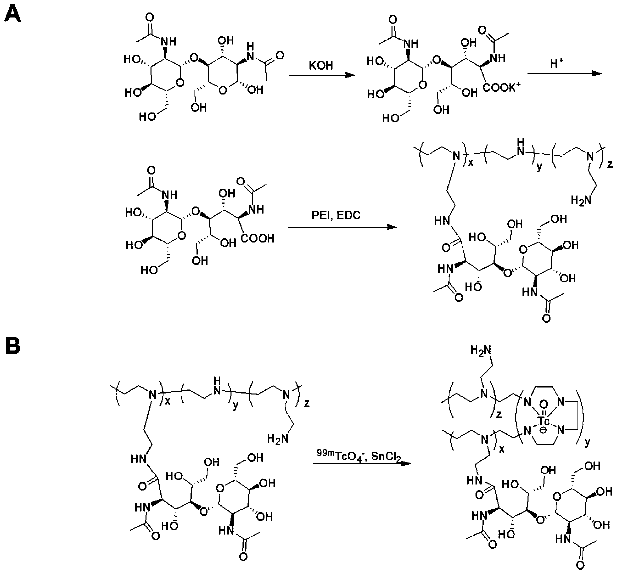
[0038] The preparation of embodiment 1 NAG-PEI and 99m Tc tag

[0039] 1. Synthesis of NAG-PEI

[0040] At room temperature, 0.1 g of chitobiose was dissolved in a mixed solution of water and methanol. Dissolve 0.285g of iodine in 4mL of methanol, dissolve and mix with the above-mentioned chitobiose. A 4% KOH solution was added to the solution until the iodine color disappeared. The solution was recrystallized with ether to obtain potassium chitobioconate. The product is obtained chitobionic acid [(NAG) by IR-120 ion exchange column 2 -COOH]. Chitobionic acid was linked to PEI (molecular weight: 1800) with EDC as a catalyst. Briefly, 25% molar ratio of chitobionic acid [(NAG) 2 -COOH] was dissolved in a solution of TEMED containing EDC to react with PEI, and stirred at room temperature for 72h. The solution was dialyzed in a dialysis bag (MWCO: 2500) for 3 days, and then freeze-dried to obtain NAG-PEI. Such as figure 1 As shown in A.

[0041] 2. Characterization of ...

Embodiment 299

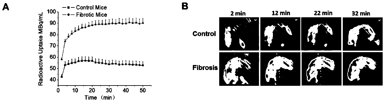
[0045] Example 2 99m Evaluation of liver fibrosis in mice by Tc-NAG-PEI SPECT / CT imaging

[0046] 1. Construction of liver fibrosis mouse model

[0047] The mice used in the experiments were female C57BL / 6 mice purchased from the Experimental Animal Center of Xiamen University. CCl 4 Used to induce liver fibrosis in mice. Simply put, CCl 4 Dissolved in olive oil (1:4), C57BL / 6 female mice were intraperitoneally injected with 5 mL / kg of CCl 4 solution, 2 times a week. The control group was given the same dose of olive oil. 5 mice in each group, 4 weeks and 8 weeks respectively.

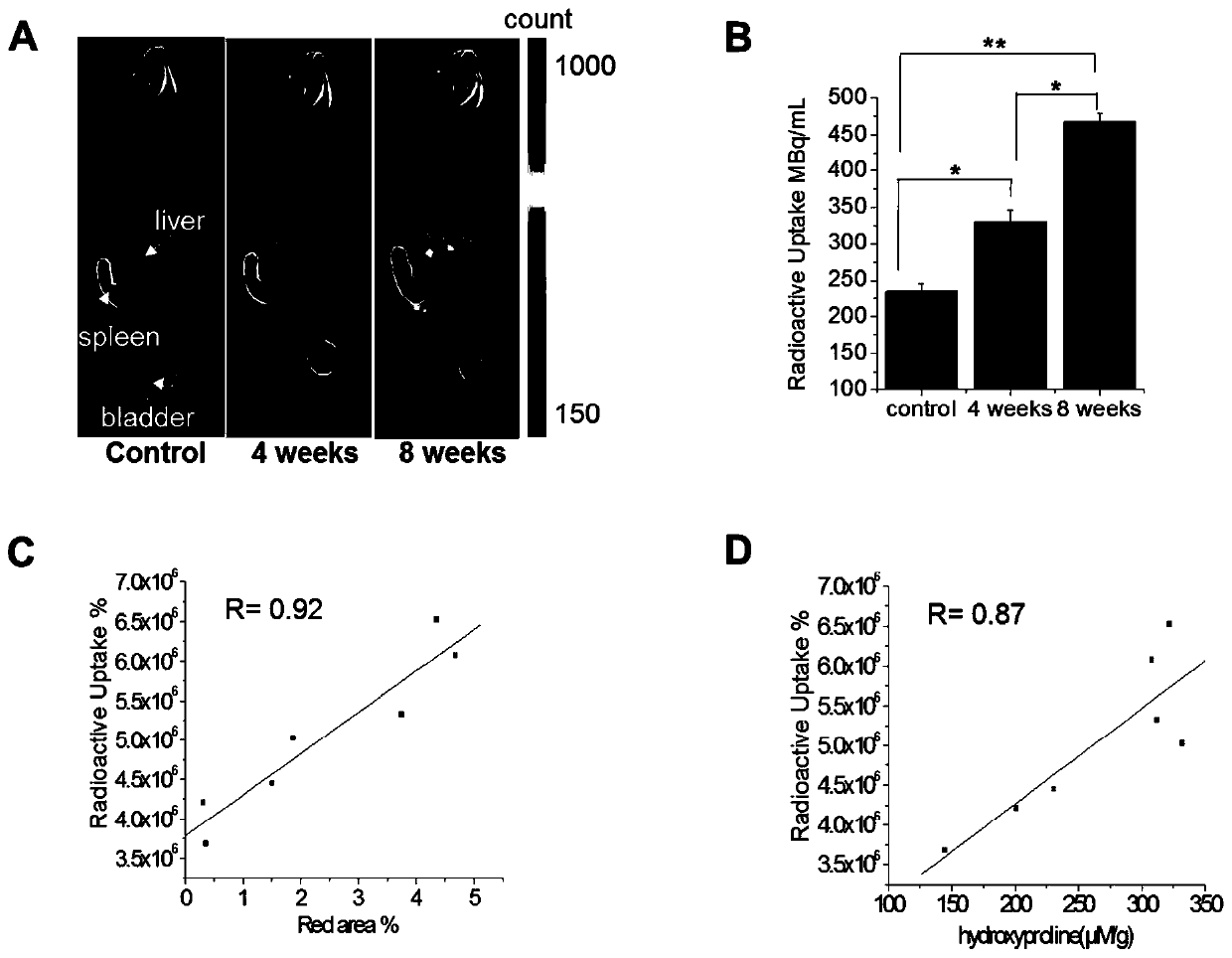
[0048] 2, 99m Tc-NAG-PEI SPECT / CT Liver Area Dynamic Scanning

[0049] The dynamic SPECT / CT scanning mice were divided into three groups: 3 mice in the control group and 3 mice in the 8-week fibrosis group. The mice were first anesthetized with isoflurane, and then placed in SPECT / CT for scanning. The whole process was anesthetized with isoflurane, the oxygen flow rate was 0.8mL / min, and the ...

Embodiment 3

[0054] Example 3 Sirius red staining and autoradiography of fibrotic liver tissue sections

[0055] 1. Tissue section and Sirius red staining

[0056]Tissue embedding: First, the mice in the control group and the fibrosis group that had been imaged were killed by necking, and the liver was taken out. Each liver was cut into two pieces of 1.5×1.5 cm approximately square liver tissue with a razor blade. The edge of the tissue was required to be flat. Wash twice with PBS solution to remove blood cells, and then fix the liver tissue in a centrifuge tube containing 4 ml of 4% paraformaldehyde solution overnight. After washing twice with PBS solution, dehydrate with graded alcohol, with alcohol concentration ranging from 50% to 100% for 30 minutes each time. If something needs to interrupt the experiment, you can put it in 70% alcohol at 4°C overnight. After dehydration, use xylene for transparency, alcohol: xylene 1:1 mixture solution for 30 minutes, then clear twice in xylene, 4...


PUM

PropertyMeasurementUnit
molecular weightaaaaaaaaaa

Abstract

The invention discloses a nuclide-labeled targeted probe compound as well as a preparation method and application thereof. The molecular weight of the nuclide-labeled targeted probe compound is 1kDa-50kDa, and the nuclide-labeled targeted probe compound comprises a polyethyleneimine skeleton and a targeted group derived from N-acetyl glucosamine or diacetyl chitobiose acid and is labeled through coordination of a radionuclide X label and an imino group on the polyethyleneimine skeleton. The nuclide-labeled targeted probe compound can be applied to in-vivo hepatic fibrosis detection, has the advantages of relatively short imaging time, relatively high imaging quality, relatively wide imaging depth, less kidney radioactive accumulation and the like, and is relatively beneficial to early hepatic fibrosis. Meanwhile, by combining with CT imaging, accurate anatomical structure signals and functional information can be obtained, thereby being beneficial to the evaluation and accurate staging of hepatic fibrosis.

Description

technical field [0001] The invention belongs to the technical field of medical imaging, and in particular relates to a nuclide-labeled targeting probe compound and a preparation method thereof. Background technique [0002] Liver fibrosis is caused by repeated liver injury and is accompanied by deposition of extracellular matrix (mainly collagen). In damaged liver tissue, extracellular matrix proteins can replace necrotic tissue by forming fibrous scars, eventually leading to liver structural disorder and abnormal liver function. Activation and proliferation of hepatic stellate cells play a major role in the pathogenesis of liver fibrosis. Activated hepatic stellate cells are characterized by accelerated proliferation, overexpression of α-smooth muscle actin (a-SMA), continuous synthesis of extracellular matrix proteins, and secretion of chemokines and profibrokines. Therefore, imaging of activated hepatic stellate cells is a good strategy to assess liver fibrosis. [000...

Claims


Application Information

Patent Timeline
Patent Type & Authority Patents(China)
IPC IPC(8): C07H5/06C07H1/00C07B59/00C09K11/06A61K51/06
CPCA61K51/065C07B59/005C07B2200/05C07H1/00C07H5/06C09K11/06C09K2211/188
Inventor 张现忠张德良庄荣强郭志德
Owner XIAMEN UNIV
Why Eureka
Try Eureka