Tissue engineering nanofiber intravascular stent and preparation method thereof

A nanofiber and tissue engineering technology, applied in the field of tissue engineering nanofiber vascular stent and its preparation, can solve the problems of postoperative embolism, intimal hyperplasia, and long-term patency reduction, so as to improve anticoagulant performance and promote adhesion , Promote the effect of transportation and discharge of metabolic waste

Active Publication Date: 2017-10-27
上海双申医疗器械股份有限公司
View PDF6 Cites 5 Cited by
  • Summary
  • Abstract
  • Description
  • Claims
  • Application Information

AI Technical Summary

Problems solved by technology

At present, autologous blood vessels and artificial blood vessels are the most widely used grafts, but both have their own advantages and disadvantages. The number of autologous blood vessels (such as great saphenous vein or internal mammary artery) is limited, and about 30% of patients lack suitable grafts. Autologous blood vessels, making it difficult to meet clinical needs
While the effect of artificial blood vessels is better in large-diameter blood vessel transplantation, complications such as intimal hyperplasia, postoperative embolism, calcification, and infection are prone to occur in small-caliber blood vessel (inner diameter less than 6mm) transplantation, resulting in a decrease in long-term patency rate and treatment. Ineffective

Method used


Image

  • Tissue engineering nanofiber intravascular stent and preparation method thereof
  • Tissue engineering nanofiber intravascular stent and preparation method thereof

Examples


Embodiment 1

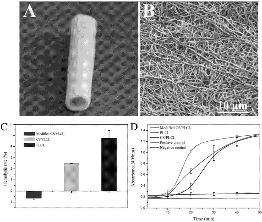
[0025] (1) Dissolve chitosan in trifluoroacetic acid / hexafluoroisopropanol solution under normal temperature conditions, stir and prepare a 6% (w / v) homogeneous solution, and dissolve PLCL in hexafluoroisopropanol under normal temperature conditions , stirred to prepare a 6% (w / v) homogeneous solution, then mixed the two evenly at a volume ratio of 1:2, and carried out electrospinning with the roller as the receiving device (electrostatic voltage: 12kV, spinning rate: 2mL / h, acceptance height: 15cm), dry the stent to remove residual solvent;

[0026] (2) Take dextran sulfate equivalent to chitosan in step (1) and dissolve it in an aqueous acetic acid solution with a mass fraction of 0.175%, and immerse the tubular stent in (1) in the above solution for 1 hour at room temperature , taking out the scaffold, washing it with deionized water, and freeze-drying to obtain the nanofiber scaffold.

Embodiment 2

[0028] (1) Dissolve polylysine in deionized water at normal temperature, stir to prepare a 6% (w / v) homogeneous solution, dissolve PLCL in hexafluoroisopropanol at normal temperature, and stir to prepare 6% (w / v) homogeneous solution, and then mix the two evenly at a volume ratio of 1:2, and use the roller as the receiving device to carry out electrospinning (electrostatic voltage: 12kV, spinning rate: 2mL / h, receiving height: 15cm), dry the bracket to remove residual solvent;

[0029] (2) Take chondroitin sulfate equal to the amount of polylysine in step (1) and dissolve it in an aqueous solution of acetic acid with a mass fraction of 0.175%. Under normal temperature conditions, immerse the tubular support in (1) in the above solution After 1 hour, the scaffold was taken out, washed with deionized water, and freeze-dried to obtain the nanofiber scaffold.

Embodiment 3

[0031] (1) Dissolve polyacrylamide in hexafluoroisopropanol at normal temperature, stir to prepare a 6% (w / v) homogeneous solution, dissolve PLCL in hexafluoroisopropanol at normal temperature, and stir to prepare 6% (w / v) homogeneous solution, and then the two were mixed uniformly at a volume ratio of 1:2, and the roller was used as a receiving device for electrospinning (electrostatic voltage: 12kV, spinning rate: 2mL / h, receiving Height: 15cm), dry the bracket to remove residual solvent;

[0032] (2) Dissolve the same amount of heparin as the polyacrylamide in step (1) in an aqueous acetic acid solution with a mass fraction of 0.175%, and immerse the tubular stent in (1) in the above solution for 1 hour at room temperature, and take out the stent , washed with deionized water, and freeze-dried to obtain the nanofiber scaffold.

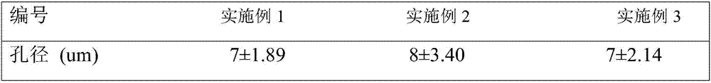
[0033] Table 1: The stent aperture value that each implementation case obtains:

[0034]


PUM

PropertyMeasurementUnit
pore sizeaaaaaaaaaa

Abstract

The invention relates to a tissue engineering nanofiber intravascular stent and a preparation method thereof. The stent consists of PLCL and a degradable polymer material, and has a nanofiber structure microcosmically, and the aperture is 5-10 microns. Preparation: dissolving the PLCL and a positively charged polymer in two different solvents to obtain homogeneous solutions, after dissolving completely, mixing two solution systems evenly, and performing electrostatic spinning by using a roller as a receiving device to obtain a nanofiber stent; and performing self-assembly modification by using a negatively charged degradable polymer as a surface modifier, washing by using deionized water, and drying to obtain a porous fiber stent. According to the tissue engineering nanofiber intravascular stent, the material is improved to improve the anticoagulant performance of the intravascular stent through performing self-assembly modification on a surface of the stent.

Description

technical field [0001] The invention belongs to the field of vascular tissue engineering scaffold and its preparation, in particular to a tissue engineering nanofiber vascular scaffold and its preparation method. Background technique [0002] With the rising incidence of cardiovascular diseases and frequent mechanical injuries, there is an urgent need for vascular grafts to repair damaged blood vessels. At present, autologous blood vessels and artificial blood vessels are the most widely used grafts, but both have their own advantages and disadvantages. The number of autologous blood vessels (such as great saphenous vein or internal mammary artery) is limited, and about 30% of patients lack suitable grafts. Autologous blood vessels make it difficult to meet clinical needs. While the effect of artificial blood vessels is better in large-diameter blood vessel transplantation, complications such as intimal hyperplasia, postoperative embolism, calcification, and infection are p...

Claims


Application Information

Patent Timeline
Patent Type & Authority Applications(China)
IPC IPC(8): A61L27/16A61L27/18A61L27/20A61L27/50A61L27/56A61L27/58
CPCA61L27/16A61L27/18A61L27/20A61L27/50A61L27/507A61L27/56A61L27/58A61L2300/42C08L67/04C08L77/04C08L33/26C08L5/08C08L5/02C08L5/10
Inventor 何创龙陶玲王伟忠杜海波刘顶华
Owner 上海双申医疗器械股份有限公司
Why Eureka
Try Eureka