

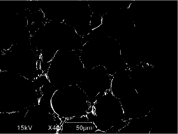
Method for preparing scanning electron microscope sample of carp muscle extracellular connective tissue
A technology for muscle cells and connective tissue, applied in the field of microscopic observation of samples, can solve problems such as unclear, incomplete scanning electron microscope photos, etc., and achieve the effect of clear structure, shortened drying time, and good tissue structure
- Summary
- Abstract
- Description
- Claims
- Application Information
AI Technical Summary
Problems solved by technology
Method used
Image

Examples
Embodiment 1
[0016] A method for preparing a scanning electron microscope sample of carp muscle extracellular connective tissue, comprising pre-fixation, post-fixation, dehydration, cutting and replacement, freeze-drying, and observation. The specific steps of preparation are:
[0017] 1) Anterior fixation: Cut carp back muscles into 5×5×10 mm pieces avoiding the diaphragm, put them in the prefixative solution for 4 days, replace the fixative solution every 24 hours, avoid the sample, and suck out the prefixation solution with a rubber dropper solution, add 1.8M NaOH solution and shake for 4 days, replace the NaOH solution every 22 hours, avoid the sample, suck out the NaOH solution with a rubber dropper, and then wash it with 0.11M phosphate buffer solution for 10 hours to obtain the pre-fixed sample. The paraformaldehyde concentration in the fixative solution is 2.1%, the glutaraldehyde concentration is 2.3%, the phosphate buffer concentration is 0.11M, and the pH of the pre-fixation solu...
Embodiment 2
[0024] A method for preparing carp muscle extracellular connective tissue scanning electron microscope samples, the specific steps of preparation are:
[0025] 1) Cut the back muscles of carp into pieces avoiding the diaphragm, put them in the pre-fixative solution for 5 days, replace the fixative solution every 23 hours, then suck out the pre-fixative solution, add a solution containing 2.1M NaOH and 0.01mM cyanuric chloride and shake for 3 days , replace the NaOH solution every 23 hours, suck out the NaOH solution, and then wash it with 0.09M phosphate buffer for 14 hours to obtain the pre-fixed sample. The buffer concentration is 0.09M, and the pH of the pre-fixation solution is 7.3. NaOH and cyanuric chloride can cooperate with each other to completely remove proteoglycans and collagen outside the connective tissue cells, and effectively reduce impurities such as extracellular matrix and elastic fibers. The impact on connective tissue, thereby improving the definition of t...
Embodiment 3
[0032] A method for preparing carp muscle extracellular connective tissue scanning electron microscope samples is as follows:
[0033] Cut the back muscles of carp into 5×5×10mm pieces avoiding the diaphragm, put them in the pre-fixation solution for 5 days, and replace the fixation solution every 24 hours. The concentration of paraformaldehyde and glutaraldehyde in the pre-fixation solution are 2% and 2.5% , The concentration of phosphate buffer solution is 0.1M, the pH of the pre-fixative solution is 7.2, then suck out the pre-fixative solution, add 2M NaOH solution, replace the NaOH solution every 24h, shake for 3 days, suck out the NaOH solution, and then wash with 0.1M phosphate buffer solution 12h, the pre-fixed sample was obtained; the pre-fixed sample was fixed in 2% tannic acid solution for 2h, then washed with 0.1M phosphate buffer for 10min, washed repeatedly for 3 times, and then washed with 1% osmium tetroxide Fix for 2 hours, wash with 0.1M phosphate buffer solut...
PUM
 Abstract
Description
Claims
Application Information