Spherical nucleic acid fluorescent probe for telomerase activity detection and preparation method and use thereof

A fluorescent probe and activity detection technology, which is applied in the field of analysis and detection and medical diagnosis, can solve the problems of low transfection efficiency and high toxicity, and achieve the effects of increasing application range, preventing agglomeration, and low cost

Active Publication Date: 2018-04-06
HEFEI INSTITUTES OF PHYSICAL SCIENCE - CHINESE ACAD OF SCI
View PDF3 Cites 8 Cited by
  • Summary
  • Abstract
  • Description
  • Claims
  • Application Information

AI Technical Summary

Problems solved by technology

Traditional transfection reagents such as liposomes and artificially modified viruses have problems such as high toxicity and low transfection efficiency, while spherical nucleic acids have a high cell uptake rate and transfection efficiency without the help of any transfection reagents. efficiency, and also protects DNA from intracellular nuclease degradation

Method used


Image

  • Spherical nucleic acid fluorescent probe for telomerase activity detection and preparation method and use thereof
  • Spherical nucleic acid fluorescent probe for telomerase activity detection and preparation method and use thereof
  • Spherical nucleic acid fluorescent probe for telomerase activity detection and preparation method and use thereof

Examples


Embodiment 1

[0055] 1. Preparation of gold nanoparticles

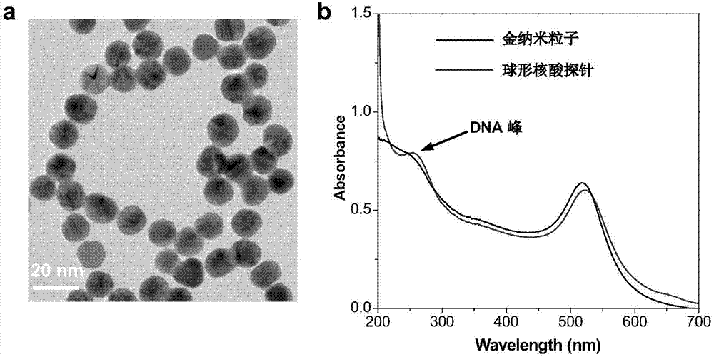
[0056] Gold nanoparticles were synthesized by reducing chloroauric acid with sodium citrate. 100mL containing 0.01wt% aqueous solution of chloroauric acid was heated to boiling, 3.5mL concentration of 1wt% sodium citrate solution was added rapidly under the situation of vigorous stirring, continued heating and reflux stirring for 10 minutes, stopped stirring, and naturally cooled to room temperature to obtain Gold nanoparticles solution (morphology see figure 2 a), store in a 4°C refrigerator for later use.

[0057] 2. Preparation of spherical nucleic acid fluorescent probes

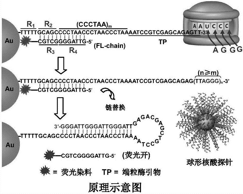
[0058] Mix the TP-chain long chain and Cy5 dye-modified FL-chain short chain at a molar ratio of 1:1, heat to 90°C for 5 minutes, then cool down to room temperature naturally to obtain hybrid double-stranded DNA; Double-stranded DNA was added to the gold nanoparticle solution prepared in step 1 at a molar ratio of 300:1, shaken at room temperature for 16 ...

Embodiment 2

[0068] Example 2: Detection of Intracellular Telomerase Activity

[0069] combine Figure 7-10, Imaging Intracellular Telomerase Activity Using Spherical Nucleic Acid Probes. Using HeLa cells as the basic model, HeLa cells were planted in confocal culture dishes and cultured for 24 hours. Then 50 μL of probe (6 nM) was added to the culture dish and incubated for 2 h, and imaged by laser confocal fluorescence microscope. In telomerase-positive HeLa cells, the FL-chain was replaced and the fluorescence recovered, so the fluorescence signal in the cytoplasm could be seen on the confocal microscopy images. The telomerase inhibitor 3′-azidovudine was co-cultured with HeLa cells for 48 hours, then 50 μL of probe was added and cultured for 2 hours. Observation with a laser confocal fluorescence microscope showed that the intracellular fluorescence signal was significantly reduced, indicating that the intracellular Telomerase activity was inhibited by telomerase inhibitors ( Figu...

Embodiment 3

[0070] Example 3: Labeling of Tumor Cells in Human Peripheral Blood

[0071] to combine Figure 11-13 , tumor cell HeLa and normal cell MRC-5 were planted in a culture dish respectively, and after culturing for 24 hours, 50 μL (6 nM) were added and cultured for 2 hours. Different proportions of HeLa and MRC-5 cells were mixed and the fluorescent signals were detected by flow cytometry. Tumor cells and normal cells doped in different proportions could be well distinguished ( Figure 11 , 12 ). On this basis, HeLa cells were planted in 20mm culture dishes and cultured for 24h. Then 50 μL of the probe was added to the dish and after 2 h of incubation, the cells were trypsinized and resuspended in 1 mL of PBS buffer. Take out 5 cells and mix them in 50 μL of human peripheral blood without red blood cells, collect the cells with a flow cytometer and detect the signal. The 5 cells mixed in the peripheral blood can be detected. In the experiment of doping 10 cells 9 ( Figure 1...


PUM


Abstract

The invention discloses a spherical nucleic acid fluorescent probe for telomerase activity detection and a preparation method and use thereof. The spherical nucleic acid fluorescent probe takes a goldnano particle as a carrier, and a double-stranded DNA is loaded on the surface of the carrier; one single-stranded DNA of the double-stranded DNA is a TP-chain long chain, the TP-chain long chain comprises a telomerase primer TP, a sequence and an auxiliary sequence complementary with a telomerase amplification sequence in turn from a 3' end; the other single-stranded DNA of the double-stranded DNA is a FL-chain short chain decorated by fluorochrome complementary with the TP-chain long chain. The spherical nucleic acid fluorescent probe overcomes heterogeneity of tumor cells, achieves universality detection of tumors on a molecular level, and can achieve cross-platform detection of the tumors from a molecule, a cell and a tissue to a living body and visual navigation for tumor surgeries.

Description

technical field [0001] The invention relates to a spherical nucleic acid fluorescent probe for detecting telomerase activity, a preparation method and application thereof, and belongs to the fields of analysis detection and medical diagnosis. Background technique [0002] Cancer is a disease of high morbidity and mortality, whose common denominator is uncontrolled cell growth. Precisely distinguishing tumor cells from normal cells in vivo is of great significance for early diagnosis, treatment and prognosis of cancer, but it is still a long-standing problem. Currently, a range of methods such as single-cell sequencing, molecular imaging, mass spectrometry, and tissue immunochemistry have been used to help resolve this difficulty. However, due to the heterogeneity of tumor cells and the lack of universal tumor markers, even the same tumor may have several to dozens of genotypes and phenotypes. Therefore, these methods are not only costly but also inaccurate. On the other ha...

Claims


Application Information

Patent Timeline
IPC IPC(8): C12Q1/6841C12Q1/6811C12Q1/48C12N15/11C12N15/10
CPCC12Q1/485C12Q1/6811C12Q1/6841G01N2333/9128C12Q2563/137C12Q2563/155C12Q2563/107
Inventor 张忠平刘正杰王振洋刘变化赵君韩光梅张瑞龙
Owner HEFEI INSTITUTES OF PHYSICAL SCIENCE - CHINESE ACAD OF SCI
Why Eureka
Try Eureka