Method for characterizing damage effect of toxin to living cells by SERS spectroscopy

A cell damage and living cell technology, applied in the fields of nanomaterials and life sciences, can solve the problem of inability to judge the degree of cell damage and apoptosis level, and achieve the effects of accurate and reliable results, simple and convenient preparation

Active Publication Date: 2018-04-20
JIANGNAN UNIV
View PDF1 Cites 3 Cited by
  • Summary
  • Abstract
  • Description
  • Claims
  • Application Information

AI Technical Summary

Problems solved by technology

[0003] In the early stage of poisoning, abrin toxin will damage the cells to a certain extent, thereby inducing cell apoptosis, and the substances and degrees of changes in cells in each apoptosis stage from apoptosis to cell necrosis are different, but only through Ordinary kits cannot judge the degree of cell damage and the level of apoptosis

Method used


Image

  • Method for characterizing damage effect of toxin to living cells by SERS spectroscopy
  • Method for characterizing damage effect of toxin to living cells by SERS spectroscopy
  • Method for characterizing damage effect of toxin to living cells by SERS spectroscopy

Examples


Embodiment 1

[0032] The synthesis route of gold seeds is as follows:

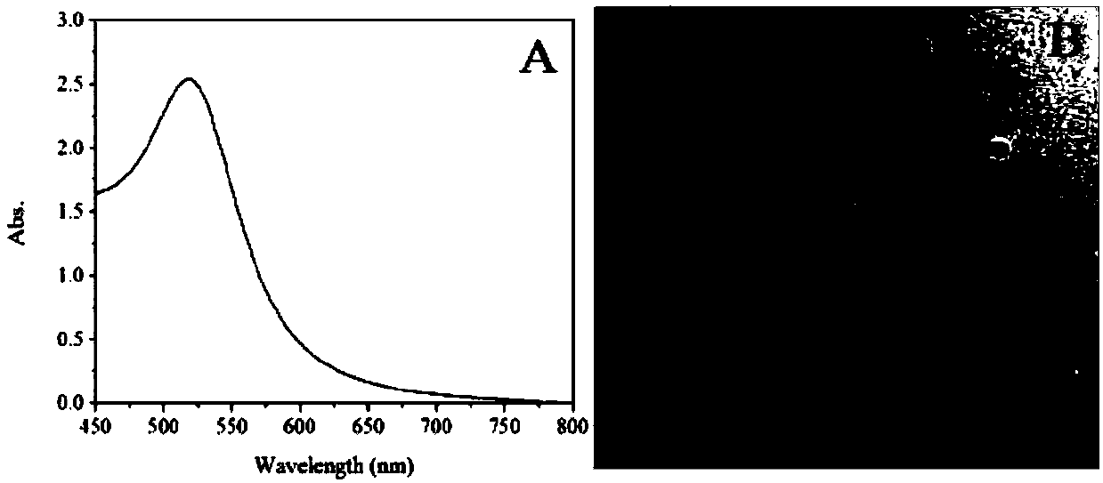
[0033] Weigh 0.2 g of trisodium citrate and dissolve it in 20 mL of ultrapure water to prepare a 1% trisodium citrate aqueous solution. 1 mL of 100 mM HAuCl 4 Add 99 mL of boiling ultrapure water. Then 15 mL of 1% trisodium citrate aqueous solution was added and vigorously stirred. After reacting for 10 minutes, the color of the gold sol turned wine red, and the reaction was terminated. The prepared gold seeds were cooled to room temperature at room temperature, and the ultraviolet-visible absorption spectrum ( figure 1 A) and TEM micrograph ( figure 1 B) Perform characterization and store at 4°C for future use.

Embodiment 2

[0035] The synthetic route of gold nanostars is as follows:

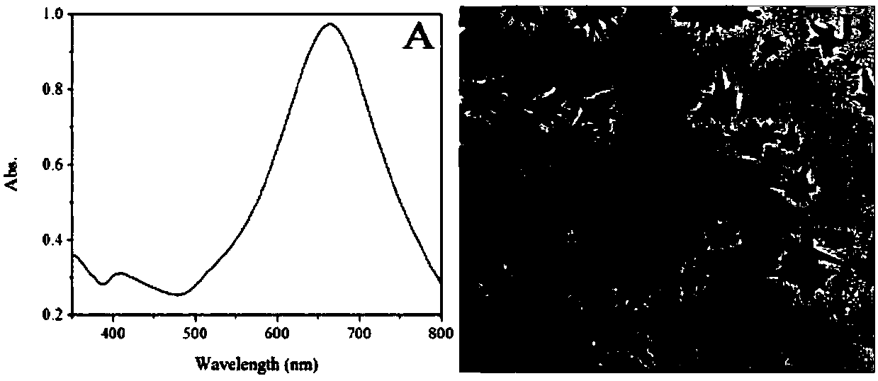
[0036] 50 μL of 100 mM HAuCl 4 The solution was added to 19.5 mL of ultrapure water that was constantly stirred, and then 200 μL of the gold seeds prepared above, 200 μL of a silver nitrate solution with a concentration of 3 mM, and 100 μL of an ascorbic acid solution with a concentration of 100 mM were added respectively, and the reaction was stirred at room temperature for 10 minutes until the color of the solution changed. It is blue-green. Finally, the reaction was terminated by centrifugation at 3000rpm for 15min, and the ultraviolet-visible absorption spectrum ( figure 2 A) and TEM micrograph ( figure 2 B) Perform characterization.

Embodiment 3

[0038] Detection of the SERS effect of gold nanostars:

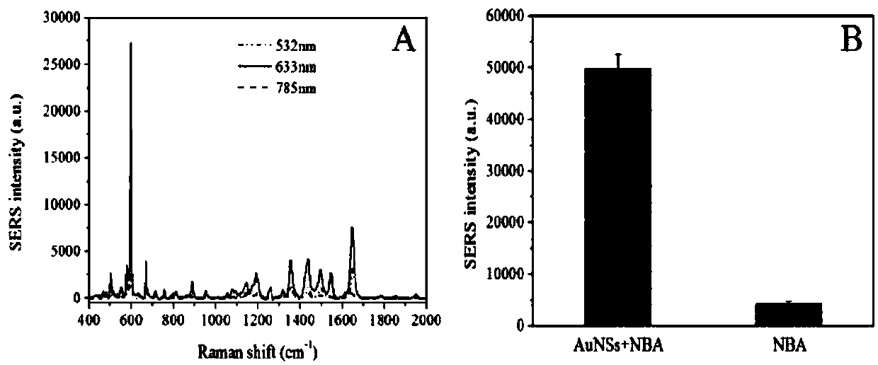
[0039]Add 200 μL of NBA aqueous solution with a concentration of 0.5 mM to 20 mL of the prepared gold nanostar solution and incubate overnight in a shaker at 37 °C. Stop the reaction with centrifugation at a speed of 3000rpm for 15min, resuspend with ultrapure water, select respectively 532nm, 633nm and 785nm wavelength diode lasers as the excitation light source, detect the SERS spectrum of NBA, and obtain the best excitation wavelength as 633nm ( image 3 A). At the excitation wavelength of 633nm, the signal intensity of the SERS spectrum of NBA measured by gold nanostars is much greater than the Raman signal of NBA at the same concentration. at 592cm -1 The NBA characteristic peak intensity at is used to compare the enhancement effect of gold nanostars ( image 3 B).


PUM


Abstract

The invention provides a method for characterizing the damage effect of toxin to living cells by SERS (surface enhanced Raman scattering) spectroscopy. Application of SERS spectroscopy to living cellcharacterization not only expands the new application of SERS spectroscopy, but also has potential application prospects in study of living cell damage. A TAT functionalized SERS probe is employed asthe Raman enhancement substrate, and enters living cells in a transmembrane way through receptor dependence mediated endocytosis, thus enhancing the Raman signal of cells themselves. The average SERSspectrum change of the cells before and after the action of toxin is detected to realize characterization of cell damage, also differential spectrum analysis and PCA are utilized to contrast the difference of cells on which the toxin acts for different periods of time, thus identifying and differentiating cells under different states. In order to further verify the cell apoptosis level, the Annexin V-FITC / PI double-staining reagent and flow cytometry are utilized to quantify the apoptosis situation. The method provided by the invention can achieve real-time nondestructive monitoring of the damage effect of toxin to living cells, and is simple and practicable, convenient and efficient.

Description

technical field [0001] The invention relates to the fields of nanomaterials and life sciences, in particular to a method for characterizing the damage effect of toxins on living cells by using SERS spectrum. Background technique [0002] Abrin, a cytotoxic protein isolated from the seeds of legumes, is more toxic than ricin, LD in mice 50 It is 0.05μg / kg, and the lethal dose for adults is 5.0-7.0μg / kg. It has been listed as a potentially important biological toxin warfare agent and one of the pathogenic substances of bioterrorism. The structure and mechanism of action of abrin toxin are similar to those of ricin. They are composed of two chains, A and B, connected by a disulfide bond. In the cell, the A chain enters the 4324th position of the 28S rRNA that catalyzes the 60S large subunit of the cell and deadenines the 60S ribosomal subunit, thereby inhibiting the synthesis of cellular proteins. Acacia toxin has no specific chemical groups and elements, and it is difficult ...

Claims


Application Information

Patent Timeline
IPC IPC(8): G01N21/65G01N15/14
CPCG01N15/14G01N21/658
Inventor 马小媛张京娜王周平
Owner JIANGNAN UNIV
Why Eureka
Try Eureka