A 3D printing bio-ink for rapid repair of spinal injury blood vessels and its preparation method

A 3D printing, spinal injury technology, applied in the field of biological 3D printing, can solve problems such as the poor application of spinal injury blood vessel repair, the difficulty of taking into account the mechanical strength and cell growth environment, cell density, and the uncontrollable space of species, etc. Conducive to growth and reproduction, avoiding cell sedimentation, and promoting rapid repair

Active Publication Date: 2021-02-05
JINAN UNIVERSITY
View PDF8 Cites 0 Cited by
  • Summary
  • Abstract
  • Description
  • Claims
  • Application Information

AI Technical Summary

Problems solved by technology

[0005] Existing bioprinting nozzles can only print one material, which is difficult to take into account factors such as mechanical strength and cell growth environment. At the same time, the cell density and type space caused by sedimentation are uncontrollable, which is not good for spinal injury vascular repair. application

Method used


Image

  • A 3D printing bio-ink for rapid repair of spinal injury blood vessels and its preparation method

Examples


Embodiment 1

[0030] (1) Preparation of shell material ink

[0031] Using deionized water as a solvent, magnetically stir 2% gelatin and 5% sodium alginate; add growth factor VEGF50ng / mL and antibacterial and anti-inflammatory drug streptomycin 100μg / mL magnetically stir evenly, and sterilize by ultraviolet light for 12 hours before use.

[0032] (2) Preparation of Inner Cell Layer Ink

[0033] At 37°C, bone marrow mesenchymal stem cells (BMSC) 1×10 6 / mL, vascular endothelial cells 1×10 6 / mL, smooth muscle fibroblasts 1×10 6 / mL, vascular endothelial growth factor (VEGF) 30ng / mL, platelet growth factor (PDGF) 30ng / mL and basophilic fibroblast growth factor (bFGF) 30ng / mL were added to the complete medium DMEM, and mixed evenly.

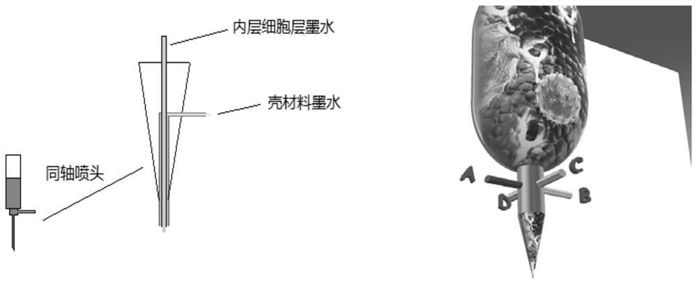
[0034] (3) Print preparation

[0035] Coaxial nozzles are used to realize simultaneous printing of shell material and inner cells. The printer adopts extrusion printing equipment. The temperature of the shell material of the printer is set at 37°C and the pr...

Embodiment 2

[0037] (1) Preparation of shell material ink

[0038] Using deionized water as a solvent, magnetically stir 6% gelatin and 2% sodium alginate evenly; add growth factor VEGF100ng / mL and antibacterial and anti-inflammatory drug streptomycin 50μg / mL magnetically stir evenly, and sterilize by ultraviolet light for 16h before use.

[0039] (2) Preparation of Inner Cell Layer Ink

[0040] At 37°C, bone marrow mesenchymal stem cells (BMSC) 3×10 6 / mL, vascular endothelial cells 3×10 6 / mL, smooth muscle fibroblasts 3×10 6 / mL, vascular endothelial growth factor (VEGF) 50ng / mL, platelet growth factor (PDGF) 50ng / mL and basophilic fibroblast growth factor (bFGF) 50ng / mL were added to the complete medium DMEM, and mixed evenly.

[0041] (3) Print preparation

[0042] Coaxial nozzles are used to realize the simultaneous printing of shell materials and inner cells. The printer adopts extrusion printing equipment. The temperature of the shell material of the printer is set at 37°C and...

Embodiment 3

[0044] (1) Preparation of shell material ink

[0045] Using deionized water as solvent, magnetically stir 5% gelatin and 2% sodium alginate evenly; add growth factor VEGF60ng / mL and antibacterial and anti-inflammatory drug streptomycin70μg / mL magnetically stir evenly, ultraviolet sterilize for 18h and set aside.

[0046] (2) Preparation of Inner Cell Layer Ink

[0047] At 37°C, bone marrow mesenchymal stem cells (BMSC) 2×10 6 / mL, vascular endothelial cells 2×10 6 / mL, smooth muscle fibroblasts 2×10 6 / mL, vascular endothelial growth factor (VEGF) 100ng / mL, platelet growth factor (PDGF) 100ng / mL and basophilic fibroblast growth factor (bFGF) 100ng / mL were added to the complete medium DMEM and mixed evenly.

[0048] (3) Print preparation

[0049] Coaxial nozzles are used to realize the simultaneous printing of shell materials and inner cells. The printer adopts extrusion printing equipment. The temperature of the shell material of the printer is set at 37°C and the printin...


PUM


Abstract

The invention discloses 3D (three-dimensional) printing bio-ink for quickly repairing injured blood vessels of vertebral columns and a method for preparing the 3D printing bio-ink, and relates to thefield of 3D bio-printing. The method includes steps of preparing ink for shell materials; preparing ink for cell layers of inner layers; carrying out 3D printing preparation and the like. The 3D printing bio-ink and the method have the advantages that 2-4 types of materials can be simultaneously printed by the 3D printing bio-ink by the aid of coaxial processes, cells of the inner layers can be effectively protected by materials for outer layers, and the problem of cell sedimentation during printing can be solved; cell types, density and distribution can be controlled, growth factors and the like are added into the 3D printing bio-ink, accordingly, quick repair on the injured blood vessels of spinal cords can be promoted, the outer layers are excellent in mechanical property, and the innerlayers are favorable for cell growth and reproduction; the shortcomings of existing bio-ink can be overcome by the aid of the 3D printing bio-ink, and 3D printing bio-printing can be effectively applied to repairing the injured blood vessels of the vertebral columns.

Description

technical field [0001] The invention relates to the field of biological 3D printing, in particular to a 3D printing bio-ink for rapid repair of spinal injury blood vessels and a preparation method thereof. Background technique [0002] Bio-3D printing technology has made outstanding contributions to solving medical problems, and its research in the fields of cell tissue culture and tissue engineering is deepening. However, bio-ink is the basis of printing, and its development needs are imminent. Armstrong, J.P. et al. developed a bioink for cartilage printing by mixing poloxamer F127, alginate, and DMEM at 4 °C, followed by cell encapsulation. Armstrong, J.P., et al."3D Bioprinting Using a Templated Porous Bioink."Advanced Healthcare Materials.14, 5, 2016. Patent CN201610269756.5 Adding cross-linked extracellular matrix nanofiber powder from pig, bovine or sheep dermis with biological macromolecules Coagulants and coagulants are used to prepare bioinks that can be used in s...

Claims


Application Information

Patent Timeline
Patent Type & Authority Patents(China)
IPC IPC(8): A61L27/20A61L27/22A61L27/38A61L27/50A61L27/54B33Y10/00B33Y70/10B33Y80/00
CPCA61L27/20A61L27/222A61L27/3804A61L27/3808A61L27/3834A61L27/3886A61L27/50A61L27/54A61L2300/406A61L2300/414A61L2430/38B33Y10/00B33Y70/00B33Y80/00C08L5/04
Inventor 薛巍宋镕光阮淼亮戴箭王永周
Owner JINAN UNIVERSITY
Why Eureka
Try Eureka