
A cartilage-bone repair scaffold with bionic gradient and its preparation method

A technology for bone repair and subchondral bone, which is used in tissue regeneration, prosthesis, medical science, etc. It can solve the problem of poor interface adhesion, inability to simulate the continuous gradient distribution characteristics, and the interface adhesion between the scaffold layer and the layer. Weak and other problems, to achieve the effect of convenient operation, good in situ induction of cartilage tissue growth, and biocompatibility in situ induction of cartilage growth

Active Publication Date: 2021-03-02
FUZHOU UNIV
View PDF13 Cites 0 Cited by
  • Summary
  • Abstract
  • Description
  • Claims
  • Application Information

AI Technical Summary

Problems solved by technology

For example, the invention patent with the application number 201810059754.2 discloses a biomimetic oriented cartilage scaffold released by two-factor programming. The two parts of the upper surface layer and the lower transitional layer prepared separately are bonded by silk fibroin to form a complete scaffold. , found that it has a good effect on promoting proliferation and differentiation of BMSCs; however, the upper and lower layers of scaffolds are combined to form an overall scaffold, which makes the interface binding force poor; the invention patent with patent number 201410397070.5 announced the integration of bionic interface structure Cartilage-bone scaffold, which is a 1:1 mixture of silk fibroin solution and hydroxyapatite aqueous solution, the main inorganic component of human bone, to prepare a calcified layer and a subchondral bone layer. The two parts of the bone bracket are well connected, which solves the problem of weak bonding between the bracket layer and the layer interface to a certain extent.
However, this method of artificially quantitatively designing the combination of hydroxyapatite particles and organic polymers directly mixed cannot simulate the continuous gradient distribution characteristics of the inorganic components of natural articular cartilage.

Method used


Image

  • A cartilage-bone repair scaffold with bionic gradient and its preparation method
  • A cartilage-bone repair scaffold with bionic gradient and its preparation method
  • A cartilage-bone repair scaffold with bionic gradient and its preparation method

Examples


Embodiment 1

[0049] 1. Preparation of cartilage layer composite precursor solution

[0050] (1) Dissolve 50 mg of sodium hyaluronate in 10 mL of deionized water, and magnetically stir for 30 minutes in a water bath at 45-60°C until completely dissolved to obtain a sodium hyaluronate aqueous solution;

[0051] (2) Dissolve 0.5 g chitosan in acetic acid solution with a volume fraction of 1%, and magnetically stir for 30 min in a water bath at 37°C to obtain chitosan acetic acid solution A;

[0052] (3) Under the condition of magnetic stirring, slowly add the sodium hyaluronate aqueous solution obtained in step (1) into the chitosan acetic acid solution A obtained in step (2), and fully stir to form a uniform mixed solution;

[0053] (4) Add 4.5 mL, 4 mg / mL of EDC and 5.6 mL, 1 mg / mL of NHS to the mixture obtained in step (3), and cross-link at room temperature for 4 h to obtain the precursor solution of the cartilage layer complex;

[0054] 2. Preparation of Subchondral Bone Layer Complex P...

Embodiment 2

[0065] 1. Preparation of cartilage layer composite precursor solution

[0066] (1) Dissolve 100 mg of sodium hyaluronate in 10 mL of deionized water, and magnetically stir for 30 minutes in a water bath at 45-60°C until completely dissolved to obtain an aqueous solution of sodium hyaluronate;

[0067] (2) Dissolve 0.75 g chitosan in an acetic acid solution with a volume fraction of 1.5%, and magnetically stir for 30 min in a water bath at 37°C to obtain chitosan acetic acid solution A;

[0068] (3) Under the condition of magnetic stirring, slowly add the sodium hyaluronate aqueous solution obtained in step (1) into the chitosan acetic acid solution A obtained in step (2), and fully stir to form a uniform mixed solution;

[0069] (4) Add 4.5 mL, 4 mg / mL of EDC and 5.6 mL, 1 mg / mL of NHS to the mixture obtained in step (3), and cross-link at room temperature for 4 h to obtain the precursor solution of the cartilage layer complex;

[0070] 2. Preparation of Subchondral Bone Laye...

Embodiment 3

[0081] 1. Preparation of cartilage layer composite precursor solution

[0082] (1) Dissolve 150 mg of sodium hyaluronate in 10 mL of deionized water, and magnetically stir for 30 minutes in a water bath at 45-60°C until completely dissolved to obtain an aqueous solution of sodium hyaluronate;

[0083] (2) Dissolve 1.0 g chitosan in acetic acid solution with a volume fraction of 2%, and magnetically stir for 30 min in a water bath at 37°C to obtain chitosan acetic acid solution A;

[0084] (3) Under the condition of magnetic stirring, slowly add the sodium hyaluronate aqueous solution obtained in step (1) into the chitosan acetic acid solution A obtained in step (2), and fully stir to form a uniform mixed solution;

[0085] (4) Add 4.5 mL, 4 mg / mL of EDC and 5.6 mL, 1 mg / mL of NHS to the mixture obtained in step (3), and cross-link at room temperature for 4 h to obtain the precursor solution of the cartilage layer complex;

[0086] 2. Preparation of Subchondral Bone Layer Comp...


PUM


Abstract

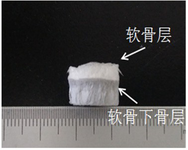
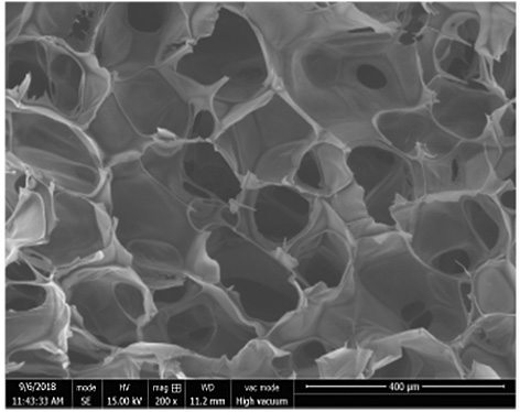
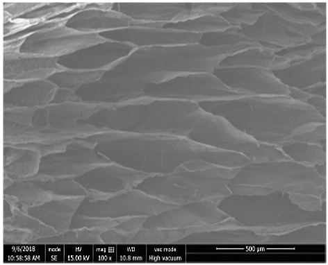
The invention discloses a cartilage-bone repair support with bionic gradient and a preparation method thereof. For the cartilage-bone repair support, a cartilage layer of an interwoven porous structure is prepared from sodium hyaluronate and chitosan, a subchondral bone layer of a porous structure is prepared from graphene oxide, chitosan and nano-hydroxyapatite, and structure, composition and function bionics of natural articular cartilage is realized by combining a coprecipitation method and a gradient refrigeration technology by referring to an in-situ bionic thorough. The obtained supporthas an upper layer of structure and a lower layer of structure, nano-hydroxyapatite presents change of bionic gradient distribution which is gradually increased from the cartilage layer to the subchondral bone layer; meanwhile, the cartilage layer in the upper layer is of a mutually penetrating interwoven hierarchical pore structure, the subchondral bone layer is of an array microtube structure which is axially arranged and is vertically through, the support material is excellent in mechanical performance, strong in interface bonding property and good in biological performance, and is expectedto become a novel composite material for treating cartilage-bone defect.

Description

technical field [0001] The invention belongs to the field of composite materials, and in particular relates to a cartilage-bone repair bracket with a bionic gradient and a preparation method thereof. Background technique [0002] Articular cartilage damage caused by trauma, disease, degeneration and other factors is a common orthopedic disease in clinical practice. Especially in the face of the aging trend of the global population, articular cartilage damage is particularly prominent. The country has brought enormous mental pressure and a heavy economic burden. Because articular chondrocytes are cells in the terminal stage of differentiation, without blood vessels, lymphatic vessels and nerves, and lack of corresponding nerve and humoral regulation, the self-repair ability of cartilage defects is limited. On the basis of traditional tissue engineering, the development of in situ tissue engineering technology provides a new opportunity for cartilage repair and functional rec...

Claims


Application Information

Patent Timeline
Patent Type & Authority Patents(China)
IPC IPC(8): A61L27/40A61L27/46A61L27/44A61L27/56
CPCA61L27/443A61L27/46A61L27/56A61L2430/06C08L5/08
Inventor 陈景帝赵耀邹琳徐敢耿玉生张其清
Owner FUZHOU UNIV
Learn More