
Magnetic nanoparticle and method for preparing elevated intraocular pressure animal model from same

A magnetic nanoparticle, animal model technology, applied in the field of nanomaterials, can solve the problems of modeling failure, short intraocular pressure time, long-term biological toxicity of tissues, etc., and achieve the effect of lasting and stable resistance

Active Publication Date: 2019-04-19
EYE & ENT HOSPITAL SHANGHAI MEDICAL SCHOOL FUDAN UNIV
View PDF9 Cites 1 Cited by
  • Summary
  • Abstract
  • Description
  • Claims
  • Application Information

AI Technical Summary

Problems solved by technology

Generally characterized by poor IOP stability and low surgical success rate
The peribulbar vein ligation can stimulate the compensatory proliferation of blood vessels, bypass the ligated vessels, and achieve venous return, resulting in the failure of modeling; the dexamethasone method can cause long-term biological toxicity to many tissues in the eye, which increases the complexity of the problem and is not conducive to the accuracy of a single problem. Investigation; the gel method maintains high intraocular pressure for a short period of time, and has limited role in the in-depth investigation of long-term high intraocular pressure diseases; the size of the magnetic beads selected by the anterior chamber magnetic bead injection method is about 3 μm, and can only be placed in the trabecular meshwork of the chamber angle Enrichment, cannot go deep into the internal tissue of the aqueous humor running channel, the basic properties of the magnetic beads used are not adjustable, and cannot be well adapted to the specific microscopic tissue structure, resulting in unsatisfactory stability and persistence of high intraocular pressure. The success rate of animal models of high intraocular pressure is less than one-third

Method used


Image

  • Magnetic nanoparticle and method for preparing elevated intraocular pressure animal model from same
  • Magnetic nanoparticle and method for preparing elevated intraocular pressure animal model from same
  • Magnetic nanoparticle and method for preparing elevated intraocular pressure animal model from same

Examples


Embodiment 1

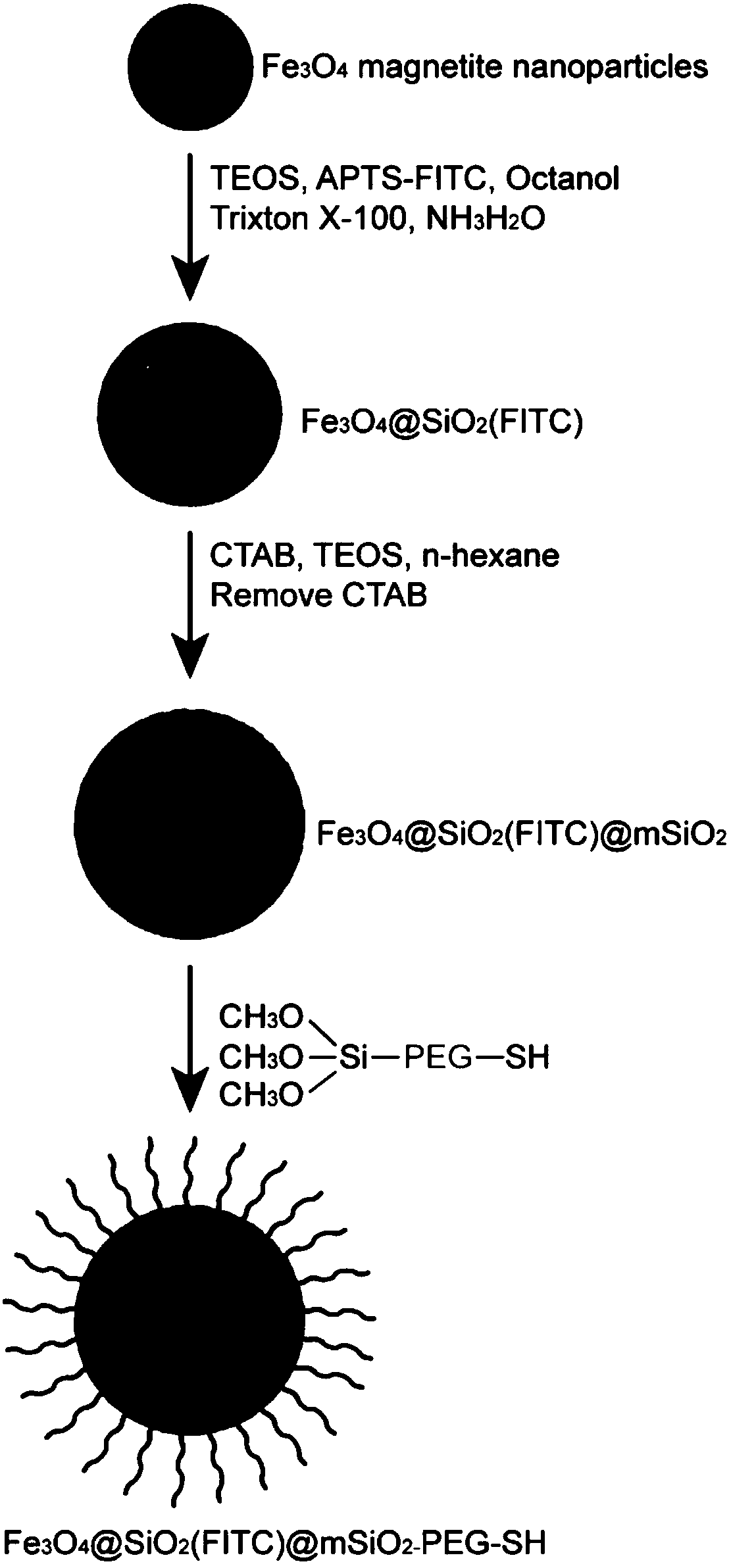
[0056] The preparation of magnetic nanoparticles adopts the following steps:

[0057] S1: Preparation of small-sized magnetic nanoparticles

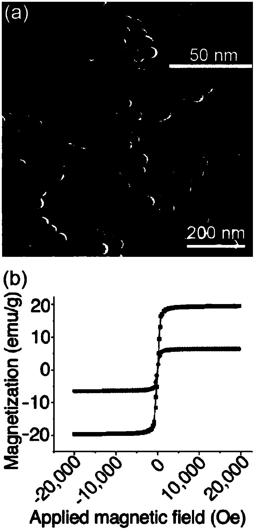
[0058] Dilute 5 mL of magnetic powder-n-hexane dispersion (20 mg / mL) in a cyclohexane solution of 10 mL of n-octanol and 10 mL of polyethylene glycol octylphenyl ether (trixton X-100). Add 0.5mL ammonia water (28wt%) to the above solution to form a stable inverse microemulsion. Then, 0.04 mL of ethyl silicate (TEOS) was added under continuous mechanical stirring, and the reaction was allowed to proceed for 24 h to form a silica-coated magnetic nanoparticle dispersion, namely Fe 3 o 4 @SiO 2 sample. Next, in the above Fe 3 o 4 @SiO 2 Fluorescent probe mother solution (0.02g 3-aminopropyltriethoxysilane (APTS), 0.01g fluorescein isothiocyanate (FITC), 0.02mL TEOS, 5mL ethanol) was added to the dispersion solution. After continuing to stir for 12 hours under dark conditions, the product was purified by magnetic separation, washed wi...

Embodiment 2

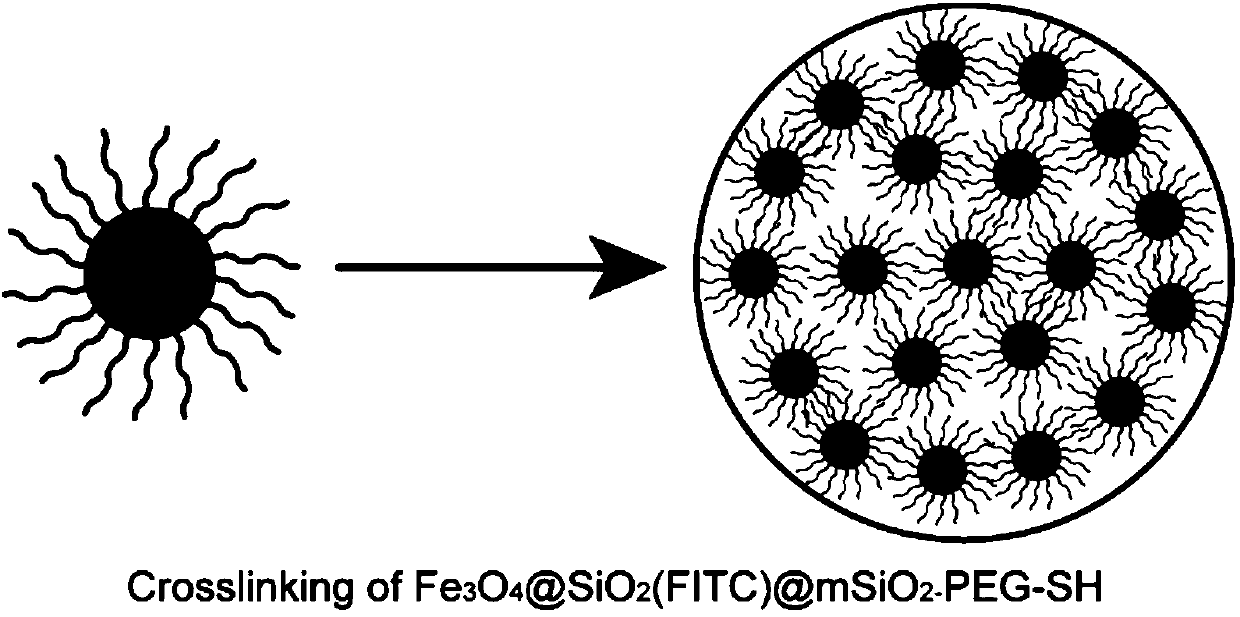
[0071] This example is basically similar to Example 1, except that the polymer molecule is polyamino acid with a molecular weight of 10,000; the particle size of small-sized magnetic nanoparticles is 50nm, and the magnetic particles formed by crosslinking small-sized nanoparticles through emulsion polymerization The particle size of the nanoparticles is 900 nm; the magnetic nanoparticles are injected into the anterior chamber in the form of an aqueous dispersion, and the dose is 80 μL. The magnetic nanoparticles are guided by a ring magnet to gather at the angle of the chamber, and gradually disperse in the trabecular meshwork, Schlemm's canal and venous capillaries as the aqueous humor circulates, and accumulate in tissues at all levels of the aqueous humor running channel. Maintain high intraocular pressure for a long time, the intraocular pressure can be increased to 4 times the normal intraocular pressure, and it can be stably maintained at a certain intraocular pressure da...

Embodiment 3

[0073] This example is basically similar to Example 1, except that the polymer molecule is polylactic acid with a molecular weight of 8000; the particle size of the small-sized magnetic nanoparticles is 70nm, and the magnetic particles formed by cross-linking the small-sized nanoparticles through emulsion polymerization The particle size of the nanoparticles is 600 nm; the magnetic nanoparticles are injected into the anterior chamber in the form of an aqueous dispersion with a dose of 60 μL. The magnetic nanoparticles are guided by a ring magnet to gather at the angle of the chamber, and gradually disperse in the trabecular meshwork, Schlemm's canal and venous capillaries as the aqueous humor circulates, and accumulate in tissues at all levels of the aqueous humor running channel. Maintain high intraocular pressure for a long time, the intraocular pressure can rise to 3 times the normal intraocular pressure, and maintain a stable intraocular pressure data interval for 8 weeks. ...


PUM

PropertyMeasurementUnit
Particle sizeaaaaaaaaaa

Abstract

The invention provides a magnetic nanoparticle and a method for preparing an elevated intraocular pressure animal model from the same. The magnetic nanoparticle is adjustable in size, is provided witha multi-stage assembly structure, and can be prepared through an emulsion polymerization method. When the elevated intraocular pressure animal model is prepared, the magnetic nanoparticle is guided into eyes through anterior chamber injection, self-crosslinking or decrosslinking can be adjusted according to a specific microstructure structure of an aqueous humor circulation path, namely a trabecular meshwork / Schlemm's canal, and a stable aqueous humor blocking layer is formed. An aqueous humor moving channel is plugged in all directions and multiple levels, aqueous humor return fluid circulation is limited, the aqueous humor outflow resistance is increased, and the elevated intraocular pressure is maintained lastingly. The preparation method of the elevated intraocular pressure animal model is simple, convenient to use, safe and long in effect.

Description

technical field [0001] The invention relates to a magnetic nano particle and a method for preparing an animal model of high intraocular pressure, belonging to the technical field of nano materials. Background technique [0002] At present, the preparation methods of animal models of glaucoma ocular hypertension include peribulbar vein ligation, dexamethasone, anterior chamber gel injection, and anterior chamber magnetic bead injection. Although these methods can prepare animal models of ocular hypertension, they have their own advantages. various problems. The general feature is poor stability of intraocular pressure and low surgical success rate. The peribulbar vein ligation can stimulate the compensatory proliferation of blood vessels, bypass the ligated vessels, and achieve venous return, resulting in the failure of modeling; the dexamethasone method can cause long-term biological toxicity to many tissues in the eye, increasing the complexity of the problem, which is not...

Claims


Application Information

Patent Timeline
IPC IPC(8): A61K41/00
CPCA61K9/0009A61K9/0019A61K41/00
Inventor 孙建国邓勇辉
Owner EYE & ENT HOSPITAL SHANGHAI MEDICAL SCHOOL FUDAN UNIV
Learn More