Luminescent ELISA in vitro diagnosis reagent kit for cerebral apoplexy and in vitro test equipment

An in vitro diagnosis and kit technology, applied in the field of biomedical testing, can solve problems such as the difficulty in distinguishing hemorrhagic stroke from ischemic stroke, the inability to judge the treatment effect of damaged cell types, and the low level of stroke diagnosis, so as to improve the accuracy and reproducibility, avoiding spinal taps, and reducing testing costs

Active Publication Date: 2019-04-30
成都蓝瑙生物技术有限公司
View PDF6 Cites 4 Cited by
  • Summary
  • Abstract
  • Description
  • Claims
  • Application Information

AI Technical Summary

Problems solved by technology

[0008] Whether it is through imaging diagnosis or ELISA cerebrospinal fluid detection, at present, the main difficulties in the diagnosis of stroke are: early asymptomatic or mild symptoms, difficult to detect; real-time diagnosis is difficult to distinguish hemorrhagic stroke from ischemic stroke; long-term treatment cannot judge the affected stroke. Impairment of cell type and / or efficacy of treatment, leading to general underdiagnosis of stroke

Method used


Image

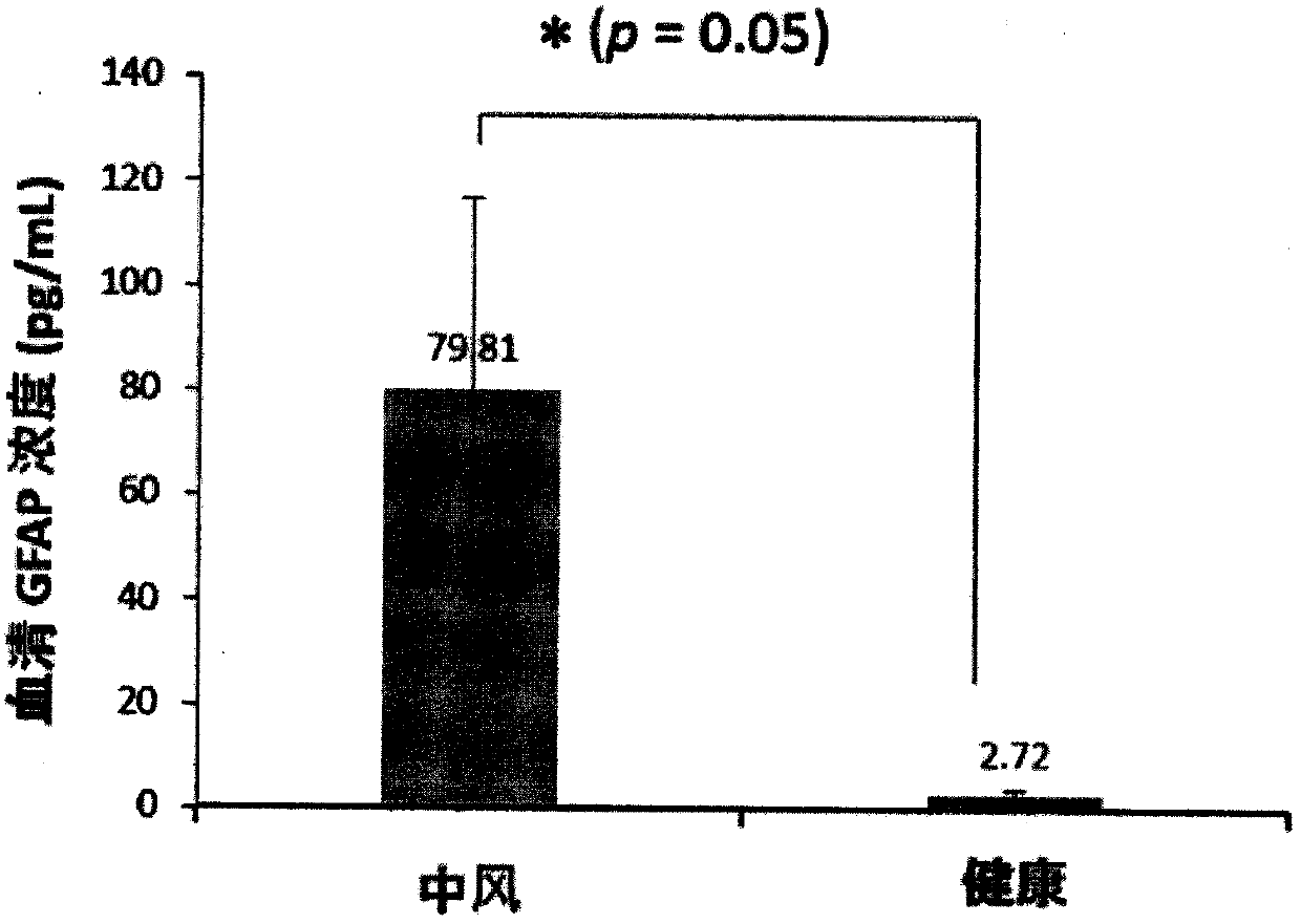
  • Luminescent ELISA in vitro diagnosis reagent kit for cerebral apoplexy and in vitro test equipment
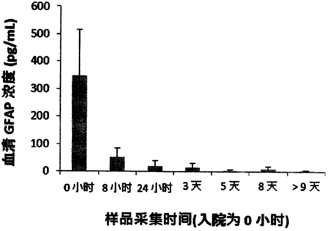
  • Luminescent ELISA in vitro diagnosis reagent kit for cerebral apoplexy and in vitro test equipment
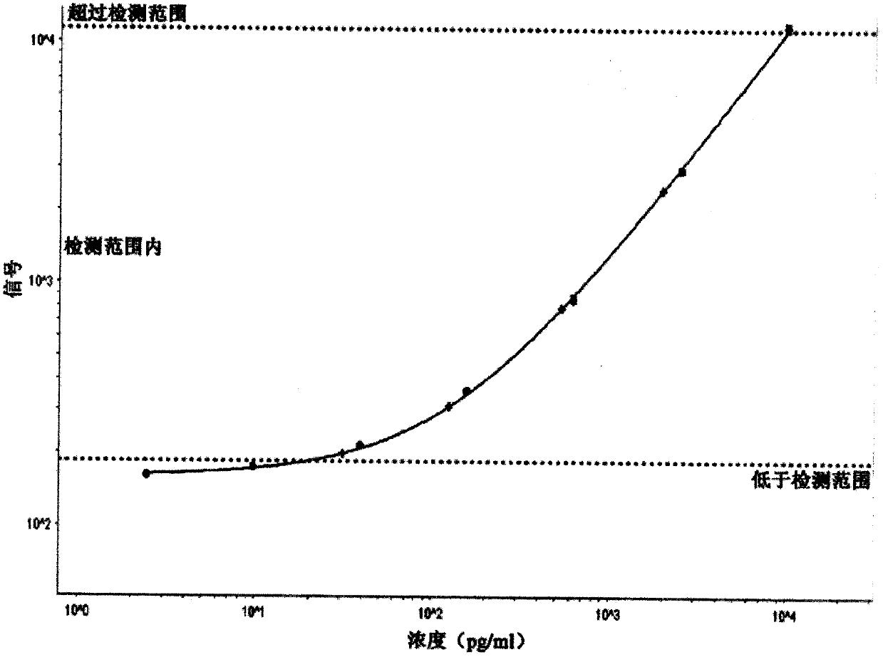
  • Luminescent ELISA in vitro diagnosis reagent kit for cerebral apoplexy and in vitro test equipment

Examples


preparation example Construction

[0074] Preparation of serum to be tested

[0075] Extract blood from cerebral apoplexy patients intravenously; place at room temperature for 1-2 hours to allow natural layering and separation of blood cells and serum; mix evenly the serum with the aqueous solution for serum sample stabilization at a ratio of 2:1-1:10, and set aside.

[0076] ELISA

[0077] Prepare 48 or 96 well microtiter plates (elisa plates). Adsorb the GFAP monoclonal antibody in the small hole of the microtiter plate, and wash once with PBS; add the diluted stroke patient serum prepared above to the small hole of the microtiter plate; then wash off the unbound excess antibody; Then, a detection antibody that has been coupled to a luminescent group in advance is added to the small well, and the coupling detection antibody is preferably an animal-derived antiserum (ie, a polyclonal antibody). Then wash the wells of the microtiter plate 3-4 times.

[0078] Antigen content determination

[0079] After wash...

preparation example 1

[0086] Preparation of Aqueous Solution 1 for Body Fluid Sample Stabilization

[0087] Prepare 50 mL of sterile distilled water, add 10 mL of normal human serum (BioreclamationIVT, USA), 1 g of bovine serum albumin (Sigma, USA), 2 mL of Tween 20 (Sigma-Aldrich, USA), and a final concentration of 0.05 mM urea (Sigma- Aldrich, USA), sodium chloride (Sigma-Aldrich, USA) at a final concentration of 110 mM, potassium chloride (Sigma-Aldrich, USA) at a final concentration of 2.0 mM, and Tris base (Sigma-Aldrich , USA), and finally add the remaining amount of sterile distilled water to mix evenly and make the volume to 100mL.

preparation example 2

[0089] Preparation of aqueous solution 2 for stabilization of body fluid samples

[0090] Prepare 50 mL of sterile distilled water, add 20 mL of normal human serum (BioreclamationIVT, USA), 2 g of bovine serum albumin (Sigma, USA), 2 mL of Tween 20 (Sigma-Aldrich, USA), and a final concentration of 0.08 mM urea (Sigma- Aldrich, USA), a final concentration of 125mM sodium chloride (Sigma-Aldrich, USA), a final concentration of 2.3mM potassium chloride (Sigma-Aldrich, USA), a final concentration of 15mM Tris base (Sigma-Aldrich, USA), and finally add the remaining amount of sterile distilled water to mix evenly and make the volume to 100mL.


PUM


Abstract

The invention provides a luminescent ELISA in vitro diagnosis reagent kit, which comprises an aqueous solution for stabilization of a body fluid sample. The body fluid sample comprises a nerve injurymarker protein component. In the aqueous solution for stabilization of the body fluid sample, solutes comprise human serum, animal serum albumin, inorganic base metal salt, Tris-methylamin, protein denaturant and nonionic surface active agent, and the pH value of the aqueous solution is 6.7-7.6. The in vitro diagnosis reagent kit optionally comprises an antibody against the nerve injury marker protein component (e.g. anti-GFAP (glial fibrillary acidic protein) antibody). The invention also provides in vitro test equipment. When a luminescent ELISA method is adopted to detect the nerve cell injury marker protein component GFAP from the body fluid sample (e.g. peripheral blood), the in vitro diagnosis reagent kit and the in vitro test equipment are very high in accuracy and repeatability, and can accurately and stably detect the pg-level GFAP in the body fluid.

Description

technical field [0001] The invention relates to the field of biomedical detection, in particular to an aqueous solution for stabilizing body fluid samples for patients with cerebral apoplexy, an in vitro detection kit, and a detection system. Background technique [0002] Stroke, commonly known as apoplexy, is caused by brain damage caused by blockage or rupture of cerebral blood vessels, and the harm is very serious. Epidemiological studies have found that the disability rate of stroke in our country ranks first, and the fatality rate ranks second, second only to cancer. There is no disease like a stroke that instantly distorts the mouth and eyes, numbs the limbs, paralyzes the bed, and makes people lose the dignity of life. At present, there are more than 3.7 million new stroke cases in my country every year, and nearly 20 million stroke patients survive, and the number of patients is in a period of rapid increase. High blood pressure, diabetes, and heart disease are all...

Claims


Application Information

Patent Timeline
Patent Type & Authority Applications(China)
IPC IPC(8): G01N33/68
CPCG01N33/6893
Inventor 邓杰丁维俊夏巍
Owner 成都蓝瑙生物技术有限公司
Why Eureka
Try Eureka