Method for improving biological mineralization capacity of silk fibroin

A technology of silk fibroin and biomineralization, which is applied in the direction of microorganism-based methods, biochemical equipment and methods, peptide preparation methods, etc., can solve the problem of silk fibroin's weak ability to induce hydroxyapatite and the limitation of silk fibrobiology The comprehensive performance of the material, the weak biomineralization function of silk fibroin, etc., achieve the effect of excellent ability to regulate the nucleation and growth of mineralization, promotion of nucleation and rapid growth, excellent water solubility and biocompatibility

Active Publication Date: 2019-10-08
ZHEJIANG UNIV
View PDF7 Cites 1 Cited by
  • Summary
  • Abstract
  • Description
  • Claims
  • Application Information

AI Technical Summary

Problems solved by technology

However, compared with sericin, which contains more acidic amino acids, the proportion of acidic amino acids in silk fibroin is less. It is generally believed that the biomineralization function of silk fibroin is weak, which limits its application in the field of biomineralization.
Studies have found that only the non-crystalline region sequence of the silk fibroin peptide chain can induce the growth of apatite crystals in the mineralization solution, and requires a longer mineralization time (7 days); while the silk fibroin peptide chain Because the crystalline region contains more non-polar amino acids, the ability to adsorb or chelate salt ions is low, so the ability to induce the formation of hydroxyapatite in the mineralization solution is very weak, so the comprehensive performance is that silk fibroin induces The ability of hydroxyapatite is very weak, even if it can induce the formation of hydroxyapatite, it needs a long mineralization time and harsh biomimetic mineralization steps
This defect limits the comprehensive performance of silk fibroin biomaterials and its application in bone tissue engineering
[0005] To sum up, there is no simple process, easy operation, and high biosafety method that can promote the biomimetic mineralization ability of silk fibroin.

Method used


Image

  • Method for improving biological mineralization capacity of silk fibroin
  • Method for improving biological mineralization capacity of silk fibroin
  • Method for improving biological mineralization capacity of silk fibroin

Examples


Embodiment 1

[0028] This embodiment includes the following steps in turn:

[0029] (1) Using phage display technology to display the amino acid sequence Thr-Lys-Arg-Glu-Glu-Val-Asp (such as SEQ ID NO.1) of the amelogenin core polypeptide fragment with the function of mineralization regulation on the P3 capsid of the phage On the protein surface, phage nanofibers exhibiting the function of inducing mineralization were prepared, called amelogenin-type phage;

[0030] (2) Add the amelogenin-type bacteriophage in step 1) and 10 mL of Escherichia coli expanded and shaken overnight into 500 mL of LB liquid medium and incubate at 37° C. for 24 hours. Then use 4500g high-speed centrifugation to collect the supernatant to remove Escherichia coli. Then use the PEG / NaCl mixed solution to precipitate the phage, spend 4 nights overnight, then centrifuge at 8000g to collect the precipitate to obtain the enriched amelogenin-type phage particles, repeat the precipitation-centrifugation step twice, so tha...

Embodiment 2

[0033] This embodiment includes the following steps in turn:

[0034] (1) Use phage display technology to display the amino acid sequence Glu-Glu–Glu-Glu-Glu-Glu-Glu-Glu-Glu (such as SEQ ID NO.2) of the mineralization-regulating dentin protein core polypeptide fragment on the P6 of the phage On the surface of the capsid protein, phage nanofibers exhibiting the function of inducing mineralization were prepared, called dentin-type phage;

[0035] (2) Add the dentin-type bacteriophage in step 1) and 5 mL of Escherichia coli expanded and shaken overnight into 100 mL of LB liquid medium and incubate at 37° C. for 12 hours. Then use 4500g high-speed centrifugation to collect the supernatant to remove Escherichia coli. Then use the PEG / NaCl mixed solution to precipitate the phage, spend 4 nights overnight, then centrifuge at 8000g to collect the precipitate to obtain the enriched dentin-type phage particles, repeat the precipitation-centrifugation step 2 times, so that the purity of...

Embodiment 3

[0038] This embodiment includes the following steps in turn:

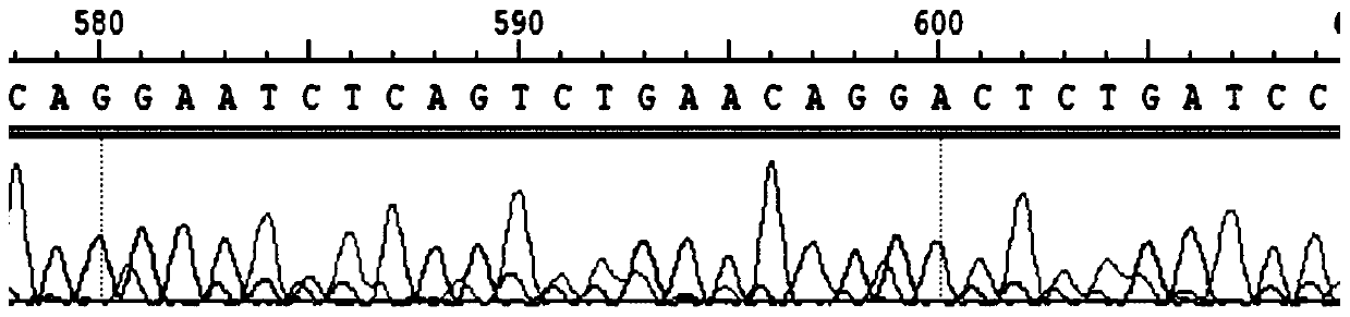
[0039] (1) Utilizing phage display technology to extract the amino acid base sequence Gln-Glu-Ser-Gln-Ser-Glu-Gln-Asp-Ser (such as SEQ ID NO.3, sequence For sequencing results, see image 3) was displayed on the surface of the P8 capsid protein of the phage, and a phage nanofiber exhibiting the function of inducing mineralization was prepared, which was called dentin matrix protein type 1 phage, and the sequencing results were as follows figure 1 shown;

[0040] (2) Add the dentin-type bacteriophage in step 1) and 4 mL of Escherichia coli expanded and shaken overnight into 100 mL of LB liquid medium and incubate at 37° C. for 16 hours. Then use 4500g high-speed centrifugation to collect the supernatant to remove Escherichia coli. Then use the PEG / NaCl mixed solution to precipitate the phage, spend 4 nights, and then centrifuge at 8000g to collect the precipitate to obtain the enriched dentin matrix protein type ...


PUM


Abstract

The invention discloses a method for improving biological mineralization capacity of silk fibroin. The method comprises the steps of through a bacteriophage displaying technique, displaying polypeptide having a mineralization adjusting and controlling function on the surface of capsid protein of bacteriophage, preparing bacteriophage nanofibers displaying a mineralization inducing function, and forming a mineralization type bacteriophage solution; performing amplification, performing purification and concentrating the mineralization type bacteriophage solution; and adding the mineralization type bacteriophage solution to a silk fibroin solution for assembly so as to obtain modified silk fibroin. The modified silk fibroin obtained by the method disclosed by the invention has more excellentcapacity for adjusting and controlling nucleation and growth of a mineralizer than silk fibroin which is not improved, the modifying method is simple to operate and effective, the modifying technologyis efficient, the production cycle is greatly shortened, and the method has the advantage that the modified silk fibroin can be obtained in a large-scale manner.

Description

technical field [0001] The invention relates to a processing method of biological materials, in particular to a method for improving the biomineralization ability of silk fibroin. Background technique [0002] Bone is the hardest connective tissue in the animal body. It is mainly composed of inorganic mineral hydroxyapatite (HA) crystals and organic type I collagen fibers and other hard tissues that are formed through the mineralization process in vivo. Biomineralization is a common life phenomenon. During the mineralization process in vivo, mineralization regulatory proteins such as osteopontin, osteocalcin, and dentin matrix protein play an important role in the crystal orientation, size, and morphology of HA crystals. control role. [0003] Bone defects and bone injuries are common diseases, with up to tens of millions of patients every year, posing a serious threat to patients' normal life activities and physical and mental health. The repair of bone tissue injury is o...

Claims


Application Information

Patent Timeline
Patent Type & Authority Applications(China)
IPC IPC(8): C07K14/435C07K1/00C12P21/00C12R1/91
CPCC07K14/43586C12P21/00
Inventor 杨明英帅亚俊段博王捷
Owner ZHEJIANG UNIV
Why Eureka
Try Eureka