Method for detecting enzymatic activity concentration through combined use of subject and object probes and nanopore technique

A host-guest, active technology, applied in the field of supramolecular chemistry, can solve the problems of strong background interference, low signal characteristics, unfavorable blocking signals and high-sensitivity detection.

Inactive Publication Date: 2019-12-24
INST OF CHEM CHINESE ACAD OF SCI
View PDF1 Cites 2 Cited by
  • Summary
  • Abstract
  • Description
  • Claims
  • Application Information

AI Technical Summary

Problems solved by technology

However, clogged signals are very unfavorable for high-sensitivity detection because of their low signal characteristics and strong background interference

Method used


Image

  • Method for detecting enzymatic activity concentration through combined use of subject and object probes and nanopore technique
  • Method for detecting enzymatic activity concentration through combined use of subject and object probes and nanopore technique
  • Method for detecting enzymatic activity concentration through combined use of subject and object probes and nanopore technique

Examples


Embodiment 1D

[0063] Example 1 Preparation of DNA-peptide molecular probe

[0064] In this example, two DNA-peptide molecular probes were prepared, and the used DNA molecules and peptide molecules were all commissioned by Sangong Bioengineering (Shanghai) to synthesize. The sequence of the DNA molecule is 5'-CATATTACACTCTCAC GACTC-3' (SEQ ID NO: 1), and its 5'end is modified with sulfhydryl groups; the sequence of peptide molecule 1 is LFG K (SEQ ID NO: 2), and the sequence of peptide molecule 2 For FGVCitK (SEQID NO: 3), the side chain of the lysine residue at the carboxyl end of the peptide molecule is modified with 3-maleimidopropionic acid.

[0065] Take 100 μL DNA (100M) and 100 μL peptide molecule 1 (5mM), mix the two evenly and adjust the pH to about 8.0 with ammonia; vortex at room temperature for 4h. The reaction solution was separated and purified by preparative anion exchange chromatography to obtain the desired DNA-peptide molecule 1, which was named as DNA-KGFL probe.

[0066] Take ...

Embodiment 2

[0067] Example 2 Construction of sample cell system

[0068] The sample cell system includes two compartments (cis and trans), the volume of both compartments is 1.0mL, and the two compartments are separated by a 20μm thick PTFE membrane (the middle of the membrane has a diameter of about 100- 150μM hole), use a glass capillary to drop a mixed solution consisting of 1 volume part of n-hexadecane and 10 volume parts of n-pentane into the pores of the polytetrafluoroethylene film (the amount of dripping is about 2 to 3 μL), Then add electrolyte buffer (3M KCl, 10mM Tris, pH 5.0) and 1,2-diphytanyl lecithin (lipids) (1,2-diphytanyl lecithin with phospholipid stock solution) to the two compartments The concentration of the phospholipid stock solution is 10 mg / mL, and the addition amount is about 15 μL; the addition amount of the electrolyte buffer is 1 mL), and mixed with a pipette to make it self-assemble to form a phospholipid bilayer. Then add 1μL of α-hemolysin protein solution ...

Embodiment 3

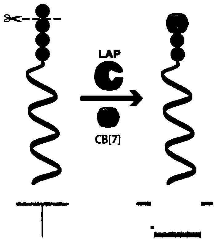
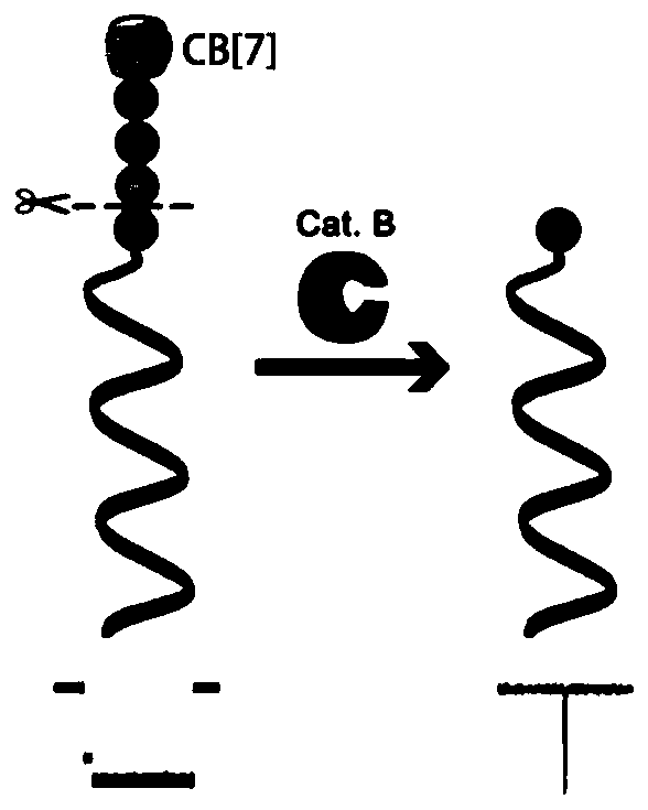
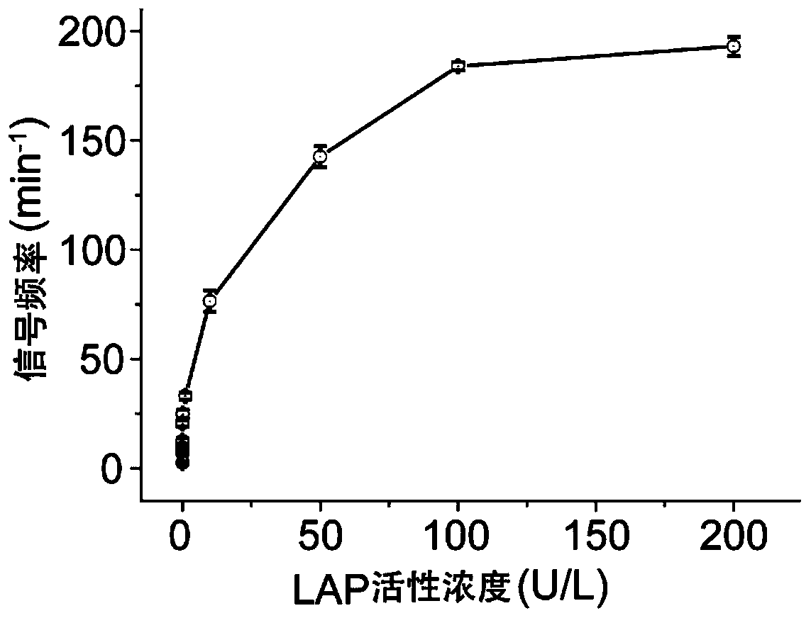
[0072] Example 3 Enzyme activity concentration detection of leucine aminopeptidase

[0073] In this example, the enzyme activity concentration of Leucine aminopeptidase (LAP) was detected. LAP was purchased from Sigma-Aldrich, 10 U / mg.

[0074] (1) Take the DNA-KGFL probe (1.23μL, 163.9μM) prepared in Example 1, and then take various concentrations of LAP and mix them in a buffer (10mM PB, pH 7.4), and adjust the total volume to 20μL, so that the final concentration of the LAP to be tested is 3.1×10 -6 , 3.1×10 -5 , 3.1×10 -4 , 3.1×10 -3 , 3.1×10 -2 , 0.31, 3.1, 15.6, 31.2, 46.8, 62.4nM; incubate together at 37°C for 3h, then add 10μL of cucurbit[7]urea (5.0mM), and incubate at 4°C for 2h to prepare the test solution.

[0075] (2) Add the test solution obtained in step (1) to the cis compartment of the sample cell prepared in Example 2, and use a patch clamp amplifier (Axopatch 200B; Axon instruments, Foster City at room temperature of 25±2°C). ,CA) for signal acquisition, the signa...


PUM

PropertyMeasurementUnit
thicknessaaaaaaaaaa

Abstract

The invention relates to a method for detecting enzymatic activity concentration through combined use of subject and object probes and a nanopore technique. The invention firstly provides a molecule combination for detecting the enzymatic activity concentration. The molecule combination comprises DNA-peptide molecules and cucurbituril, wherein the DNA-peptide molecules consist of double-stranded DNA molecules and peptide molecules, the carboxyl terminals of the peptide molecules are in covalent connection to 5' terminals or 3' terminals of the double-stranded DNA molecules, and after the DNA-peptide molecules are subjected to action by enzymes to be detected, the exposure state of amino terminal aromatic amino acids is changed. After the enzymes to be detected excise a part of peptide chains through an enzymatic reaction, exposed (or lost) aromatic amino acids can (cannot) generate subject and object action with the cucurbituril, and the DNA subject and object probes can (cannot) be formed. When penetrating through nanopores, the DNA subject and object probes can produce characteristic current signals, and through detecting the current signals, high-sensitivity high-specificity detection of the enzymatic activity concentration can be realized.

Description

Technical field [0001] The invention relates to the technical field of supramolecular chemistry, in particular to a method for detecting the concentration of enzyme activity combined with host-guest probes and nanopore technology. Background technique [0002] As a class of important biomarkers, the rapid and sensitive detection of protease activity is not only of great significance for the early diagnosis of tumors, but also an important means for the observation of therapeutic effects, tumor staging and prognosis. Fluorescence spectroscopy is currently the most common enzyme activity concentration detection technology. Although the fluorescence method has high sensitivity and good spatial resolution, fluorescent probes usually require complex design and background interference caused by autofluorescence will cause the signal-to-noise ratio to decrease and affect the sensitivity and imaging quality of the method. In recent years, with the rapid development of nanotechnology, va...

Claims


Application Information

Patent Timeline
Patent Type & Authority Applications(China)
IPC IPC(8): C12Q1/37G01N27/26C12N15/11
CPCC12Q1/37G01N27/26
Inventor 吴海臣郭秉元
Owner INST OF CHEM CHINESE ACAD OF SCI
Why Eureka
Try Eureka