Electrochemical immunosensor for measuring secretory autophagosome and preparation method and application thereof

An immunosensor and autophagosome technology, applied in the field of biomedical detection, can solve the problem of fast, simple, and direct detection of secreted autophagosomes.

Active Publication Date: 2019-12-31
SOUTHEAST UNIV
View PDF8 Cites 2 Cited by
  • Summary
  • Abstract
  • Description
  • Claims
  • Application Information

AI Technical Summary

Problems solved by technology

[0004] At present, the existing methods for detecting autophagy include direct observation by transmission electron microscope, GFP-LC3 tracer, western blot detection of autophagy-related genes, and immunohistochemical detection of LC3B, etc., but there is no rapid, simple and direct detection of secreted autophagy cells. body method

Method used


Image

  • Electrochemical immunosensor for measuring secretory autophagosome and preparation method and application thereof
  • Electrochemical immunosensor for measuring secretory autophagosome and preparation method and application thereof
  • Electrochemical immunosensor for measuring secretory autophagosome and preparation method and application thereof

Examples


Embodiment 1

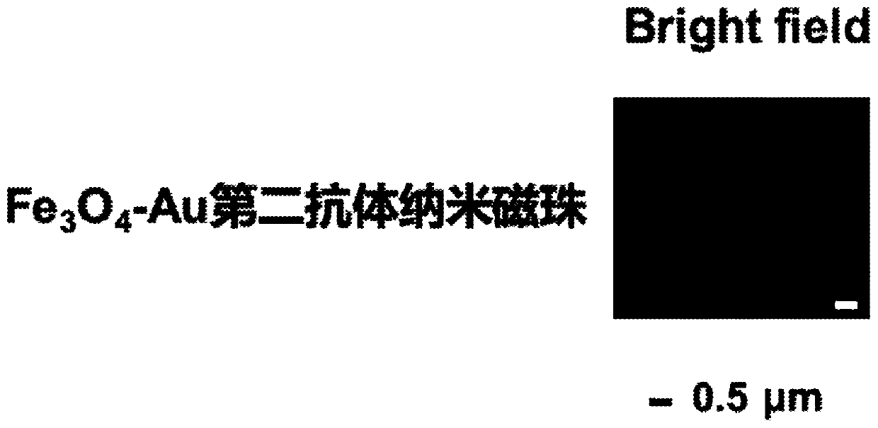
[0058] Example 1: Fe 3 o 4 - Preparation of Au Secondary Antibody Nanomagnetic Beads

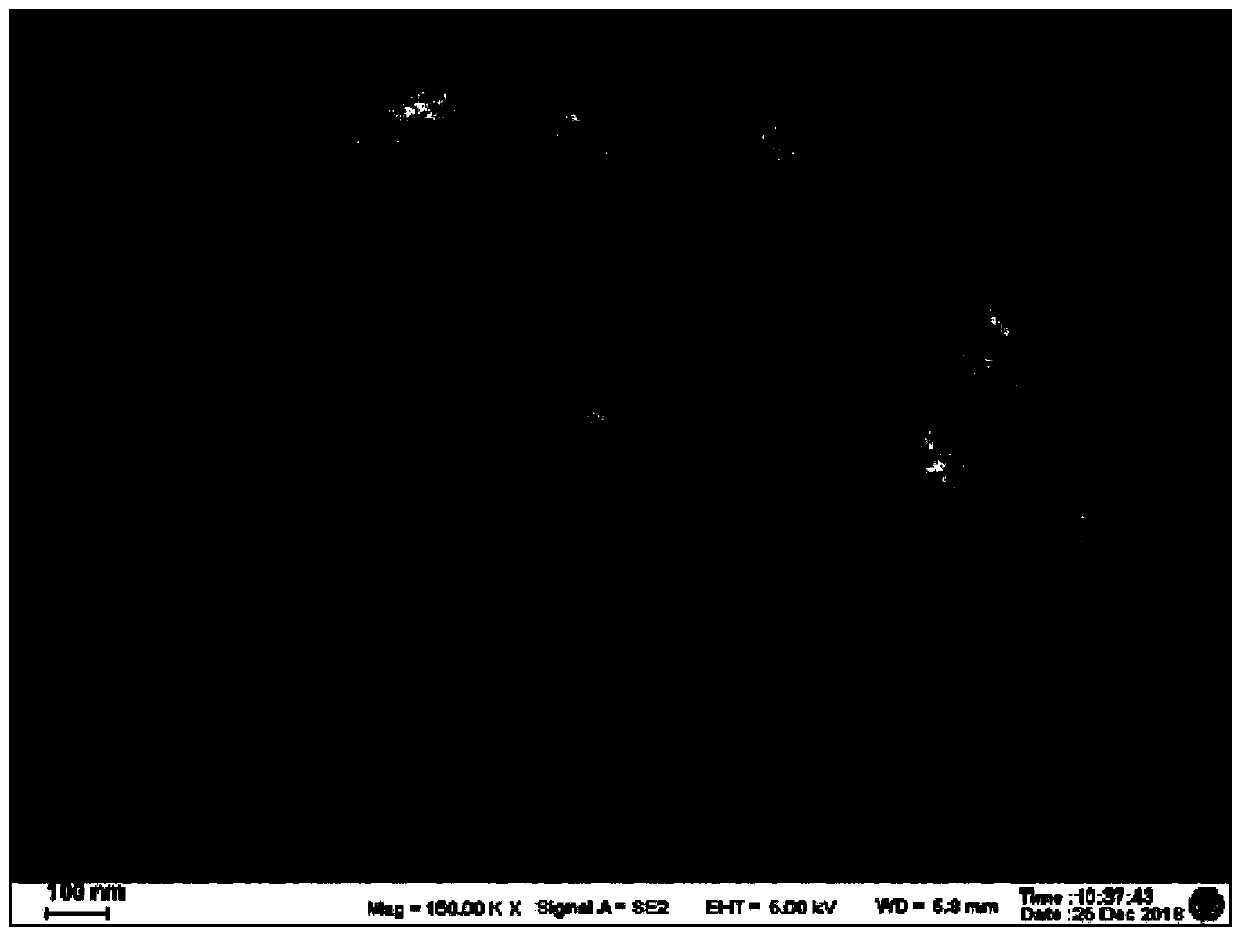
[0059] (1) Fe 3 o 4 preparation of

[0060] 0.65g FeCl 3 Dissolve 0.2g of sodium citrate in 20mL of ethylene glycol, then add 1.2g of sodium acetate, stir vigorously for 30 minutes, pour into a hydrothermal reactor, and react at 200°C for 10 hours. After the end of the reaction, the material is centrifuged to obtain the first precipitate. After the first precipitate is washed with water, it is centrifuged to obtain the second precipitate, and after the second precipitate is washed with ethanol, a liquid dissolved in ethanol is obtained to obtain Fe 3 o 4 (Concentration is 1mg / mL, pH value is 9.5), and its scanning electron microscope picture is as follows figure 1 shown.

[0061] (2) Preparation of AuNPs

[0062] 100mL mass fraction 0.01% HAuCl 4 The aqueous solution was heated to boiling, and 2 mL of a 1% sodium citrate aqueous solution was added with stirring, and continued heati...

Embodiment 2

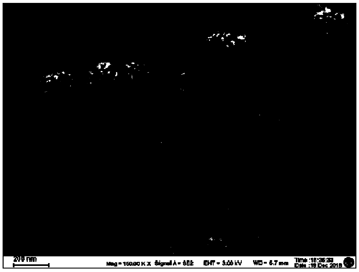
[0067] Example 2: Fe 3 o 4 -Specific verification of Au secondary antibody nano-magnetic beads

[0068] Fe 3 o 4 - Au secondary antibody nano-magnetic beads and interference red fluorescent protein co-incubated at room temperature for 2 hours, another group of Fe 3 o 4 -Au secondary antibody nano-magnetic beads were incubated with red fluorescent protein and TRAP stained with green fluorescence for 2 hours at room temperature, magnetically separated, washed 3 times in PBS, and resuspended in PBS. It was found that nano-magnetic beads can specifically capture TRAP, but not Non-specific adsorption of interfering proteins. Laser confocal images such as Figure 4 shown.

Embodiment 3

[0069] Example 3: Preparation method and detection method of electrochemical immunosensor for measuring secreted autophagosomes

[0070] The preparation method of electrochemical immunosensor is as follows: Figure 5 shown, including the following steps:

[0071] (1) Electrode pretreatment: use 0.05 and 0.03 μm Al on glassy carbon electrodes respectively 2 o 3 Powder treatment, and then ultrasonic cleaning with absolute ethanol and ultrapure water for 5 minutes;

[0072] (2) Adding GH-MB dropwise: Take 10 μL of 1 mg / mL GH-MB solution dropwise on the surface of the pretreated glassy carbon electrode with a pipette gun, place it at 37°C for 2 hours to dry, wash the unbound composite material with PBS solution, and let it dry in the air. Dry, take advantage of its larger specific surface area to enrich more antibodies on the electrode surface;

[0073] (3) Covalently link the primary antibody to the capture antibody LC3B: Add 10 μL of DC / NHS solution (20 mg / mL, 10 mg / mL) drop...


PUM

PropertyMeasurementUnit
concentrationaaaaaaaaaa

Abstract

The invention discloses an electrochemical immunosensor for measuring a secretory autophagosome as well as a preparation method and an application of the electrochemical immunosensor, and belongs to the technical field of biomedical detection. A substrate electrode is modified by a GH-MB solution, a surface marker LC3B first antibody solution of the autophagosome is covalently bound to the modified substrate electrode, and the electrochemical immunosensor is prepared. The invention further provides a method for detecting the secretory autophagosome in human peripheral blood and mouse peripheral blood by using the electrochemical immunosensor based on GH-MB signal amplification. The secretory autophagosome is captured by a Fe3O4-Au modified LC3B second antibody and then specifically bound to the LC3B first antibody. The detection method is high in sensitivity, good in specificity, wide in linear range, low in detection limit and low in cost.

Description

technical field [0001] The invention belongs to the technical field of biomedical detection, and in particular relates to an electrochemical immunosensor for detecting secreted autophagosomes and a preparation method and application thereof. Background technique [0002] Tumor is a disease with high morbidity and high mortality that seriously threatens human health. A large number of studies and clinical data have confirmed that early diagnosis and early treatment are the most effective ways to prevent and treat cancer and reduce the mortality of cancer patients. Therefore, finding tumor markers that are helpful for early diagnosis has been the focus of people's attention. Tumor markers (tumor markers, TM) refer to a class that is synthesized and secreted by the gene expression of tumor cells during the occurrence and proliferation of malignant tumors, or abnormally produced and / or increased by the body's response to tumors. These substances can provide effective informatio...

Claims


Application Information

Patent Timeline
Patent Type & Authority Applications(China)
IPC IPC(8): G01N27/327G01N33/68
CPCG01N27/3275G01N33/6854
Inventor 王立新沈艳飞张越
Owner SOUTHEAST UNIV
Why Eureka
Try Eureka