Method for separating umbilical cord mesenchymal stem cells

A technology for stem cells and separation methods, applied in cell dissociation methods, animal cells, vertebrate cells, etc., can solve the problems of aging, loss of cell phenotype, and long cell culture cycle.

Pending Publication Date: 2020-02-18
武汉济源高科技有限公司
View PDF6 Cites 1 Cited by
  • Summary
  • Abstract
  • Description
  • Claims
  • Application Information

AI Technical Summary

Problems solved by technology

The adherence method has high purity and is easier to operate than the enzymatic digestion method, but it takes a long time for cells to crawl out, the overall cell culture cycle is long, and the culture efficiency is not high; while the enzymatic digestion method is short in time, but the operation is complicated and the purity of the cells obtained by enzymatic digestion is low , and inevitably cause cell damage and loss of cell phenotype, aging

Method used


Image

  • Method for separating umbilical cord mesenchymal stem cells
  • Method for separating umbilical cord mesenchymal stem cells

Examples


Embodiment approach 1

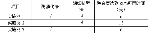
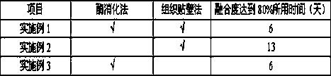
[0012] The combination of enzymatic digestion and adherence method to separate umbilical cord mesenchymal stem cells is also the method of the present invention, and the specific steps are as follows:

[0013] (1) Coating of the culture bottle: one day before the umbilical cord collection, one 75cm 2 For NUNC culture flasks, the specific coating method is as follows: dissolve 25ug of fibronectin, 25ug of laminin, and 50ug of gelatin in 5ml of stem cell culture medium, and then add them to the bottom area of ​​75cm 2 In the culture flask, the final concentration of fibronectin, laminin and gelatin were 5ug / ml, 5ug / ml and 10ug / ml respectively. Shake well, cover tightly and place in a 4°C refrigerator overnight.

[0014] (2) Mixed enzyme digestion: Break off the tip of the 10ml pipette, take 2ml of the tissue to be used in a 15ml centrifuge tube, add the corresponding volume of 0.2% hyaluronidase and 0.4% type II collagenase mixed digestion solution 2ml, shake well, place in a ...

Embodiment example 2

[0017] The pure tissue adherence method is used to isolate umbilical cord mesenchymal stem cells, and the specific steps are as follows:

[0018] 1) Adhesive culture of tissue pieces: take the above tissue pieces to be used, use a 10ml pipette with a broken tip, and pipette 2ml of tissue pieces into an uncoated 75cm 2 In the NUNC culture bottle, fully disperse the tissue pieces with 10ml of stem cell culture medium, and then culture them in a 37°C, 5% carbon dioxide incubator.

[0019] 2) Observation of results: Observe the tissue blocks and cells climbing out of the culture flask every day. It is found that on the first day, there are basically no adherent tissue blocks. On the fifth day, only 50%-70% of the tissue blocks are tightly attached to the wall. A large number of tissues floated, and 10ml full volume of liquid was changed on the 5th day, which caused a little adherent tissue to float. On the 9th day, a small amount of cells crawled out around the tissue block, and ...

Embodiment example 3

[0021] Simple enzymatic hydrolysis method to isolate umbilical cord mesenchymal stem cells, the specific steps are as follows:

[0022] (1) Mixed enzyme digestion: Break off the tip of the 10ml pipette, take 2ml of the tissue to be used in a 15ml centrifuge tube, add the corresponding volume of 0.2% hyaluronidase and 0.4% type II collagenase mixed digestion solution 2ml, shake well and place in a carbon dioxide incubator at 37°C, digest until the tissue pieces appear fluffy, then filter through an 80-mesh sieve, wash the filtrate with 5 times the volume of stem cell culture medium, centrifuge at 1500rpm for 5min, and finally Wash once with 5ml of stem cell medium, and centrifuge at 1500rpm for 5min.

[0023] (2) Cell culture: The cells at the bottom are resuspended with 10ml of new stem cell culture medium, mixed with a pipette, and then transferred to an uncoated 75cm 2 Put it in a NUNC culture bottle, and then put it into a 37°C, 5% carbon dioxide incubator for culture. Obs...


PUM


Abstract

The invention discloses a method for separating umbilical cord mesenchymal stem cells. The method specifically comprises the following steps: in a umbilical cord tissue block adherent culture process,cutting fresh Wharton's jelly umbilical cord tissue blocks into pieces with the size of about 1 mm <3>, primarily dissociating and softening the pieces with collagenase and hyaluronidase, and then uniformly spreading the pieces in a culture vessel coated with fibronectin, laminin, collagen and the like for pretreatment. By means of the method, the tissue blocks are tightly adhered, few floating tissue blocks exist, the mesenchymal stem cells in the umbilical cords can rapidly climb out of parenchyma of the tissue blocks, the cell climbing-out time is greatly shortened, meanwhile, the number of the tissue blocks climbing out of the cells is large, the amount of harvested cells is large, and the cell purity and activity are good.

Description

technical field [0001] The invention relates to a method for separating cells by adhering to tissue blocks, and more particularly to a method for separating umbilical cord mesenchymal stem cells, belonging to the field of cell biology. Background technique [0002] Umbilical cord mesenchymal stem cells exist in large quantities in umbilical cord Wharton's jelly (Wharton's jelly, or also known as Walton's jelly) and perivascular tissue, and have the potential of self-renewal, proliferation and multidirectional differentiation. At present, mesenchymal stem cells have become a hot spot in regenerative medicine research and have a very broad application prospect. [0003] The umbilical cord of newborns is rich in umbilical cord mesenchymal stem cells, and its surroundings are rich in matrix components such as collagen, hyaluronic acid, and chondroitin sulfate. There are currently two methods for the isolation and culture of umbilical cord mesenchymal stem cells, namely, the tra...

Claims


Application Information

Patent Timeline
IPC IPC(8): C12N5/0775
CPCC12N5/0665C12N2509/00
Inventor 孔德照陈杰
Owner 武汉济源高科技有限公司
Why Eureka
Try Eureka