Microporous composite biological ceramic bone repair scaffold as well as preparation method and device thereof

A technology for composite bioceramics and bone repair, applied in medical science, prosthesis, additive processing, etc., can solve the problems of large differences in natural bone, reduce wear, improve differentiation and osteogenesis efficiency, and improve spatial structure and biological performance

Pending Publication Date: 2020-09-08
BEIJING STOMATOLOGY HOSPITAL CAPITAL MEDICAL UNIV
View PDF0 Cites 5 Cited by
  • Summary
  • Abstract
  • Description
  • Claims
  • Application Information

AI Technical Summary

Problems solved by technology

[0007] It can be seen from Table 1 that the current single-component ceramic materials are quite different from human natural bone in terms of biomechanical properties, such as the index elastic modulus, fracture toughness, compressive strength and tensile strength in the table.

Method used


Image

  • Microporous composite biological ceramic bone repair scaffold as well as preparation method and device thereof
  • Microporous composite biological ceramic bone repair scaffold as well as preparation method and device thereof
  • Microporous composite biological ceramic bone repair scaffold as well as preparation method and device thereof

Examples


preparation example Construction

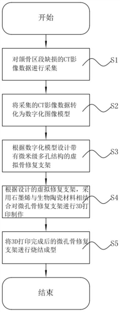
[0063] Such as figure 1 Shown, this preparation method comprises:

[0064] S1: Collect CT image data of jaw segmental defects; the data can be spiral CT or CBCT imaging data, and the scanning slice thickness is 0.2-0.625mm, which is convenient for obtaining clear CT impact data.

[0065] S2: Transform the collected CT image data into a digital image model; use the currently commonly used commercial image processing software to establish a digital three-dimensional image model of the jaw defect area, and display the shape and thickness of soft and hard tissues in the bone defect area, etc. Through the indicators such as the shape and thickness of soft and hard tissues, it is convenient to carry out data analysis on the digital image model.

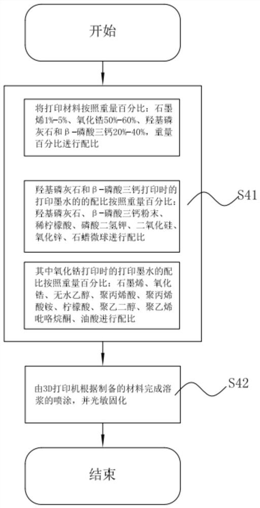
[0066] S3: Design a virtual bone repair scaffold with a micron-scale porous structure according to the digital model; at the same time, design a virtual bone repair scaffold with a suitable pore size and porosity according to the precisio...

Embodiment 1

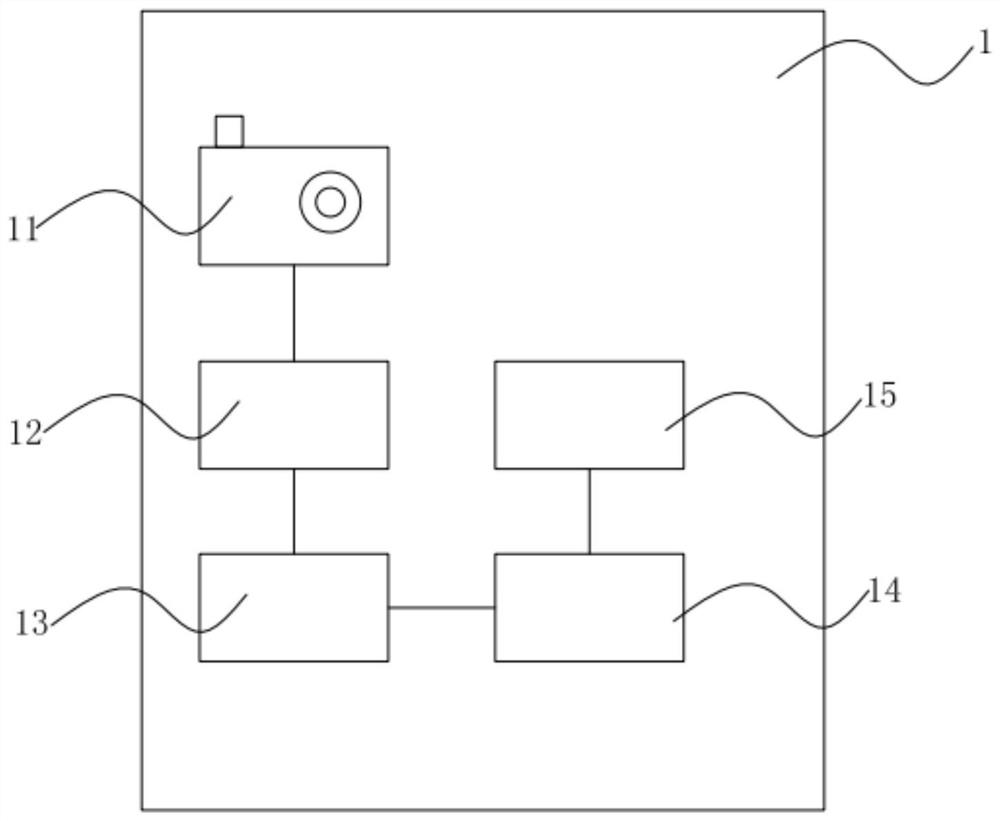
[0089] Digital design of the microporous bone repair bracket: collect the spiral CT or CBCT imaging data of the jaw segmental defect through the data acquisition part 11, and the scanning layer thickness is 0.425mm, and clear imaging data can be obtained, which is convenient for data conversion and image Processing software integration, using data conversion component 12 (commercialized image processing software) to establish a digital three-dimensional image model of the jaw defect area, respectively displaying the shape and thickness of soft and hard tissues in the bone defect area, and aiming at image distortion after digital three-dimensional imaging or noise interference, denoising processing is carried out by means of filtering, and two image quality evaluation standards, signal mean square error (MSE) and peak signal-to-noise ratio (PSNR) are applied, and the reconstructed image after denoising and the original image are evaluated by mean square Error (MSE) and peak sign...

Embodiment 2-3

[0093] As shown in Table 2, the difference from Example 1 is: the weight percentage of each component of the printing material and the printing ink component weight percentage of hydroxyapatite, β-tricalcium phosphate and zirconia are different, and other technical solutions are different from Example 1 is the same.

[0094]

[0095] Table 2: Comparison table of the weight percentages of the components of the 3D printing materials, hydroxyapatite and β-tricalcium phosphate printing inks, and zirconia printing inks of Examples 1-3

[0096] In summary, by adjusting the proportion of graphene and bioceramic materials, the conductivity and high toughness of graphene materials, the high strength and wear resistance of zirconia, and the use of hydroxyapatite and β-phosphate The good osteoinductive activity of tricalcium makes the physical properties of the new material, such as compressive strength, elastic modulus, fracture toughness and other indicators, closer to natural bone ...


PUM

PropertyMeasurementUnit
porosityaaaaaaaaaa

Abstract

The invention discloses a microporous composite biological ceramic bone repair scaffold and a preparation method and device thereof. The repair scaffold comprises a repair scaffold body made of a composite material, the repair scaffold body comprises a plurality of scaffold layers which are overlapped, a plurality of micron-sized micropores are formed in each scaffold layer, and a plurality of micropores among the layers form a plurality of polyhedrons. The composite material comprises the following components in percentage by weight: 1%-5% of graphene, 50%-60% of zirconium oxide and 20%-40% of hydroxyapatite and beta-tricalcium phosphate. The polyhedrons are connected by the micropores, the conductivity and the high toughness of the graphene are utilized, the high strength and the wear resistance of the zirconium oxide are utilized, and the good bone induction activity of the hydroxyapatite and beta-tricalcium phosphate is utilized, so that the physical properties such as compressivestrength, elastic modulus, fracture toughness and other indexes of the new material are close to natural bone tissue, the new material can bear the large stress deformation without fracture, the wearcondition between the material and the adjacent tissue is reduced, and the new material has high osteogenesis induction activity at the same time.

Description

technical field [0001] The invention relates to the field of oral and maxillofacial bone defect repair, in particular to a microporous composite bioceramic bone repair bracket and a preparation method and device thereof. Background technique [0002] The current bone defect repair techniques are as follows: [0003] Repair methods for jaw defects: such as Figure 8 As shown, diseases such as oral and maxillofacial trauma, tumors, congenital deformities, and infections often lead to partial or even overall defects of maxillofacial bone tissue. At present, there are a series of difficult problems in the treatment of patients with bone tissue defects in the oral and maxillofacial region, especially those with large-scale bone defects. Accompanied by facial tissue displacement, deformity, poor bone union, nonunion, bone exposure and other problems. In current clinical practice, such as Figure 9 In (a) and (b), Figure 10 As shown, large maxillofacial bone tissue defects are...

Claims


Application Information

Patent Timeline
Patent Type & Authority Applications(China)
IPC IPC(8): A61L27/56A61L27/08A61L27/02A61L27/12A61L27/50B33Y70/00B33Y10/00B33Y30/00
CPCA61L27/025A61L27/08A61L27/12A61L27/50A61L27/56B33Y10/00B33Y30/00B33Y70/00
Inventor 张凯宇田凯月李怀学张晓彤马晓辉王琳陈仁吉
Owner BEIJING STOMATOLOGY HOSPITAL CAPITAL MEDICAL UNIV
Why Eureka
Try Eureka