Fluorescent microsphere detection device for gastric function and gastric cancer risk and preparation method thereof

A technology of fluorescent microspheres and detection devices, which can be used in measuring devices, biological testing, material inspection products, etc. It can solve the problems of easy quenching of fluorescent antibodies, insufficient amount of protein adsorbed by NC membranes, and weak binding force, etc., to improve sensitivity , Accurate early warning and disease risk judgment, the effect of reducing dosage

Active Publication Date: 2020-09-08
JILIN PROVINCE GERUISITE BIOTECHNOLOGY CO LTD
View PDF8 Cites 4 Cited by
  • Summary
  • Abstract
  • Description
  • Claims
  • Application Information

AI Technical Summary

Problems solved by technology

[0010] The invention provides a fluorescent microsphere detection device for gastric function and gastric cancer risk and its preparation method to solve the problems in the prior art that the fluorescent antibody is easily quenched, the amount of protein adsorbed by the NC membrane is insufficient, and the binding force is not strong

Method used


Image

  • Fluorescent microsphere detection device for gastric function and gastric cancer risk and preparation method thereof
  • Fluorescent microsphere detection device for gastric function and gastric cancer risk and preparation method thereof
  • Fluorescent microsphere detection device for gastric function and gastric cancer risk and preparation method thereof

Examples


Embodiment 1

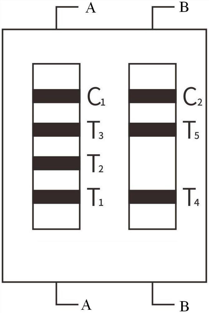
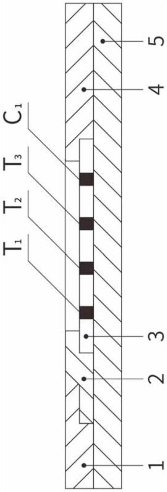
[0044] The sample pad 1, the immunofluorescent antibody glass fiber membrane 2, the nitrocellulose membrane 3, and the absorbent pad 4 are respectively pasted on the plastic plate 5, and the two ends of the nitrocellulose membrane 3 are connected to the absorbent pad 4, the immunofluorescent antibody glass fiber membrane, respectively. 2 overlapping, the other end of the immunofluorescence antibody glass fiber membrane 2 is overlapped with the sample pad 1; the nitrocellulose membrane 3 is provided with a first detection line T1, a second detection line T2, a third detection line T3, The fourth detection line T4, the fifth detection line T5, the quality control line (C1) and the quality control line (C2); the solid phase of the first detection line T1 has a highly specific gastrin 17 antibody, and the The solid phase of the second detection line T2 has a highly specific pepsinogen I antibody, the solid phase of the third detection line T3 has a highly specific pepsinogen II ant...

Embodiment 2

[0064] Preparation of polyethylene glycol glycerin treatment solution: mixed with polyethylene glycol glycerin and polylysine (SIGMA, 150KD), wherein the concentration of polyethylene glycol glycerin is 0.5%, and polylysine The concentration is 0.5%, filtered through a 0.22μm filter membrane, and set aside;

[0065] The concentration of Tris-HCL solution is 0.1mol / L, the concentration of bovine serum albumin BSA is 0.7%, the concentration of casein is 0.15%, and the concentration of surfactant is 0.7%;

[0066] All the other are with embodiment 1.

Embodiment 3

[0068] Preparation of polyethylene glycol glycerin treatment solution: mixed with polyethylene glycol glycerin, polylysine (SIGMA, 150KD) and PEG2000, wherein the concentration of polyethylene glycol glycerin is 0.5%, poly The concentration of lysine is 0.5%, the concentration of PEG20000 is 0.1%, and it is filtered through a 0.22 μm filter membrane, and it is set aside;

[0069] The concentration of Tris-HCL solution is 0.1 mol / L, the concentration of bovine serum albumin BSA is 1%, the concentration of casein is 0.2%, and the concentration of surfactant is 1%.

[0070] All the other are with embodiment 1.

[0071] Further illustrate the effect of the present invention by experiment below.


PUM


Abstract

The invention relates to a fluorescent microsphere detection device for gastric function and gastric cancer risk and a preparation method thereof, and belongs to the field of medical detection equipment. The device is prepared by adhering a nitrocellulose membrane with solid phase containing high-specificity gastrin 17, pepsinogen I, pepsinogen II antibody, anti-human IgG antibody, anti-human IgMantibody and goat-anti-mouse IgG polyclonal antibody, glass fibers adsorbed with fluorescent microsphere labeled gastrin 17, pepsinogen I, pepsinogen II antibodies and helicobacter pylori urease antigens, a sample pad, absorbent paper and other auxiliary materials. The reaction sensitivity is effectively improved; under the same threshold value, the dosage of the immunofluorescence microspheres can be reduced, the cost is saved; five gastric cancer risk markers including gastrin 17, pepsinogen I, pepsinogen II, helicobacter pylori urease IgG antibody and helicobacter pylori urease IgM antibodyin a specimen can be detected at the same time, and the complexity of production operation is not increased. The detection test paper is high in sensitivity, strong in specificity and strong in practicability.

Description

technical field [0001] The invention relates to the field of medical detection equipment, in particular to a combined detection device for G-17, PGI, PGII, HP urease IgG and IgM antibodies and a preparation method thereof, which uses immunofluorescence chromatography technology and the principle of competition method to quantitatively detect whole blood, Human gastrin 17 (G-17), pepsinogen I (PGI), pepsinogen II (PGII), Helicobacter pylori (HP) urease IgG antibody and Helicobacter pylori urease IgM antibody in serum and plasma specimens The content detection device and the preparation method thereof can realize gastric function evaluation and gastric cancer risk evaluation, and can realize sensitive, specific and rapid detection of various markers at the same time. Background technique [0002] Gastric mucosal lesions are caused by a variety of factors, including drugs, alcohol, abnormal gastric acid secretion, and Helicobacter pylori infection, among which Helicobacter pylo...

Claims


Application Information

Patent Timeline
Patent Type & Authority Applications(China)
IPC IPC(8): G01N33/74G01N33/573G01N33/558G01N33/58G01N33/574G01N33/543
CPCG01N33/74G01N33/573G01N33/558G01N33/582G01N33/57484G01N33/57446G01N33/54346G01N2333/595G01N2333/96477G01N2333/98G01N2800/60G01N2800/06
Inventor 杨小军李欣
Owner JILIN PROVINCE GERUISITE BIOTECHNOLOGY CO LTD
Why Eureka
Try Eureka