Cartilage repair material, and cartilage reconstruction biological scaffold and preparation method thereof

A cartilage repair and bio-scaffold technology, applied in the field of cartilage reconstruction bio-scaffolds and cartilage repair materials, can solve the problems of lack of mechanical strength, loss of collagen synthesis performance, inability to guarantee long-term and differentiation ability of chondrocytes, etc., to achieve guaranteed Effects of mechanical strength, promotion of regeneration, and maintenance of differentiation ability

Pending Publication Date: 2020-09-25
亘元(天津)生物医药科技有限公司
View PDF6 Cites 0 Cited by
  • Summary
  • Abstract
  • Description
  • Claims
  • Application Information

AI Technical Summary

Problems solved by technology

First of all, alternative materials or scaffolds lack the conduction and induction ability of cartilage, and lack certain mechanical strength; at the same time, as scaffold materials, transplanted or cultured chondrocytes cannot regain their original synthetic properties, which can be called chondrocytes (P0 ) will dedifferentiate under in vitro culture conditions, thereby losing the synthesis performance of collagen
In methods to date, the known three-dimensional structures for culturing chondrocytes cannot be specifically adapted to each cartilage defect
In addition, there is no guarantee that the transplanted chondrocytes will remain at the defect site for a long time and maintain the ability to differentiate, thereby allowing cartilage regeneration

Method used


Image

  • Cartilage repair material, and cartilage reconstruction biological scaffold and preparation method thereof
  • Cartilage repair material, and cartilage reconstruction biological scaffold and preparation method thereof
  • Cartilage repair material, and cartilage reconstruction biological scaffold and preparation method thereof

Examples


Embodiment 1

[0060] Example 1: Preparation of Rat Tail Collagen

[0061] 1.1 In a 10,000-level purification environment, soak the SD rat tail in a 75% ethanol solution at -20 degrees Celsius for 10 minutes to sterilize the epidermis. Peel off the epidermis, remove the head and tail to take the middle section, and extract the silvery white tail tendon. Add 0.05M Tris-HCL solution (pH7.5) containing 1M NaCl at a feed ratio of 1:10 and soak for 24 hours to remove excess polysaccharide and fat from the tail bond, and filter to remove the filtrate.

[0062] 1.2 Add 0.05M pre-cooled acetic acid solution to the pretreated silver-white tail tendon at a feeding ratio of 1:20, add 2% pepsin by volume, and react with shaking at 4°C for 36 hours until the solution gradually becomes clear transparent. Centrifuge at 5000g for 20min at 4°C, and take the supernatant;

[0063] 1.3 Add sodium chloride to a concentration of 2M, precipitate for 4 hours, 5000g, centrifuge for 20 minutes to remove the supern...

Embodiment 2

[0067] Example 2: Preparation of recombinant human truncated fibronectin rhFn and recombinant human truncated cartilage morphogenetic protein rhCDMF1

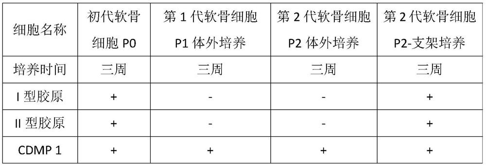
[0068] 2.1 According to the full gene sequence of human fibronectin in GenBank, the protein function and structure were analyzed, and the truncated rhFn protein sequence suitable for chondrocyte differentiation, migration and localization was reconstructed, the sequence is shown in SEQ NO: 1;

[0069] 2.2 According to the full gene sequence of human fibronectin in GenBank, the protein function and structure were analyzed, and the truncated rhCDMP1 protein sequence suitable for chondrocyte differentiation, migration and localization was reconstructed, the sequence is shown in SEQ NO: 2;

[0070] 2.3 The rhFn protein sequence and rhCDMP1 protein sequence were compiled according to the codon preference of prokaryotic cells, and the above sequences were codon-optimized and inserted into the pET28a vector, and used after prokaryotic ex...

Embodiment 3

[0073] Example 3 Preparation of Injectable Cartilage Repair Material

[0074] 3.1 Preparation of biological matrix

[0075] Take 100ml of the standard collagen solution in Example 1, quantitatively add it to the rhFN of Example 2 to a concentration of 100ug / mL, then add rhCDMP1 to a concentration of 100ng / mL; carry out aseptic subpackaging according to the actual dosage, and store at -20°C Store frozen.

[0076] 3.2 Preparation of solidification solution

[0077] Take 75ml of 2×DMEM medium and add 20% serum to mix uniformly, then add 5ml of pre-configured 25uM Hepes buffer (pH7.6), mix well, filter and sterilize, and then carry out aseptic distribution according to the actual dosage. Store frozen at -20°C.

[0078] 3.3 Directly aseptically pour the biological matrix and solidification solution into the double chamber of the prefilled syringe, tighten the sterile cap, put it in an aluminum foil blister together with the mixing chamber and the injection chamber, and store it ...


PUM

PropertyMeasurementUnit
molecular weightaaaaaaaaaa

Abstract

The invention provides a cartilage repair material, and a cartilage reconstruction biological scaffold and a preparation method thereof. The cartilage repair material comprises a biological matrix anda solidification liquid, wherein the biological matrix comprises collagen, fibronectin and cartilage morphogenetic protein, and the biological matrix, the fibronectin and the cartilage morphogeneticprotein are mixed to prepare a solution or freeze-dried powder for use; the solidification liquid comprises a DMEM culture medium, serum and a Hepes buffer solution; when the cartilage repair materialis in use, the biological matrix and the solidification liquid can be mixed and then directly filled into a cartilage defect part, and can also be solidified and formed in vitro in advance to preparethe cartilage reconstruction biological scaffold for cartilage repair. When in use, the cartilage repair material can also comprise chondrocytes or stem cells, and thereby the differentiation capacity of the cartilage repair material is further enhanced. The cartilage repair material has the beneficial effects as follows: the defects on the cartilage can be effectively filled, and the conductionand induction capabilities of the cartilage are achieved while the mechanical strength is ensured; and in addition, the cartilage repair material can keep transplanted cartilage cells at the defect part for a long time and keep the differentiation capacity of the cartilage cells, and thus promote regeneration of the cartilage cells.

Description

technical field [0001] The invention belongs to the field of tissue engineering and regenerative medicine, and in particular relates to a cartilage repair material, a cartilage reconstruction biological scaffold and a preparation method. Background technique [0002] Sports medicine is an emerging comprehensive applied discipline, mainly involving knee, shoulder, ankle, elbow and other joint injuries and osteoarthritis. Articular cartilage damage, in particular, is a common and painful condition for the patient. Damage to bone and cartilage is the pathogenesis of arthritis and is an important factor in traumatic injury and loosening of internal prostheses. Such diseases can be treated by autologous tissue or allogeneic materials and alternative materials. Autologous materials and allogeneic materials are subject to the risk of material source and infection and cannot be applied on a large scale and transformed into medicine. Therefore, alternative materials have become the...

Claims


Application Information

Patent Timeline
Patent Type & Authority Applications(China)
IPC IPC(8): A61L27/26A61L27/24A61L27/22A61L27/38A61L27/54B33Y10/00B33Y70/00B33Y80/00
CPCA61L27/227A61L27/24A61L27/26A61L27/3817A61L27/3834A61L27/54A61L2300/412A61L2430/06B33Y10/00B33Y70/00B33Y80/00C08L89/00
Inventor 王鹏
Owner 亘元(天津)生物医药科技有限公司
Why Eureka
Try Eureka