Beta-galactosidase near-infrared fluorescent probe, preparation method and application thereof

A galactosidase and fluorescent probe technology, applied in the field of biological detection, achieves the effect of stable photophysical activity and high sensitivity

Pending Publication Date: 2020-10-16
FUDAN UNIV
View PDF4 Cites 4 Cited by
  • Summary
  • Abstract
  • Description
  • Claims
  • Application Information

AI Technical Summary

Problems solved by technology

At present, a variety of β-galactosidase fluorescent probes have been reported successively, but practice has proved that the existing β-galactosidase probes are still designed based on the electronic push-pull effect after the

Method used


Image

  • Beta-galactosidase near-infrared fluorescent probe, preparation method and application thereof
  • Beta-galactosidase near-infrared fluorescent probe, preparation method and application thereof
  • Beta-galactosidase near-infrared fluorescent probe, preparation method and application thereof

Examples


Embodiment 1

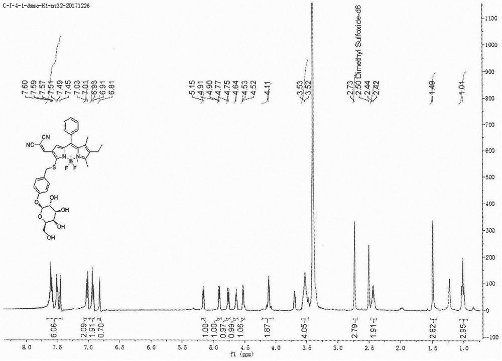
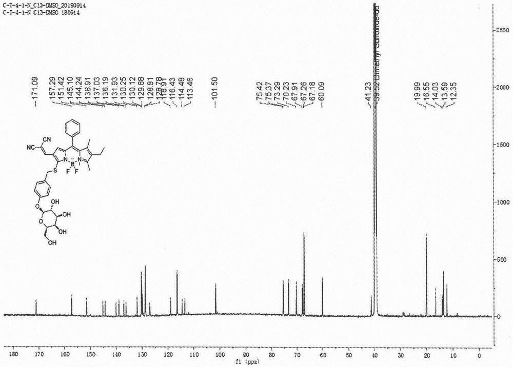
[0070] Synthetic probe general formula (1), its structure is:

[0071]

[0072] The synthesis process of the probe is as follows:

[0073]

[0074] 1) Preparation of Compound C:

[0075]

[0076] Boron trifluoride ether solution (1.8 mL, 13.2 mmol) was added to compound B (12 g, 96 mmol) in thioglycolic acid (30 mL), reacted at room temperature for 3 h, and monitored by TLC until the reaction was complete. The solution was diluted with ethyl acetate, washed with saturated brine, and the organic phase was subjected to silica gel column chromatography to obtain Compound C (14 g, 80%) as a yellow oil. 1 H NMR (400MHz, CDCl 3 ), δ2.27(s, 3H), 3.99(s, 2H), 5.66(s, 1H), 6.98(d, J=8.1Hz, 2H), 7.07(d, J=8.1Hz, 2H).ESI -MS calculated for C 9 h 10 NaO 2 S + [M+Na] + : 205.03, found: 205.1.

[0077] 2) Preparation of compound D:

[0078]

[0079] The mixture of compound C (11.7g, 64mmol) and A-D-pentaacetylgalactose (12.5g, 32mmol) was dissolved in DMF (100ml), and ...

Embodiment 2

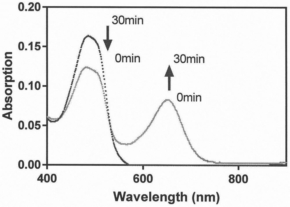
[0095] Example 2 Changes in UV absorption spectra before and after the reaction between probe C-1 and β-galactosidase

[0096] Prepare 30% DMSO in PBS solution for immediate use; prepare the probe into 1mM acetonitrile solution for later use; prepare β-galactosidase (1mg, 1580u / mg) with deionized water into 1000u / mL stock solution, aliquoted and stored at -20°C, for later use, add 3mL of 30% DMSO PBS (pH 7.4, 50mM) buffer solution to a 3mL quartz cuvette, then add 10μL of probe, 14μL of β-galactoside Enzyme, react 30min under 37 ℃, measure its absorption spectrum, by image 3 It can be seen that after the reaction of probe C-1, the maximum ultraviolet absorption intensity at 488 nm becomes weaker, and the maximum ultraviolet absorption intensity at 651 nm becomes stronger.

Embodiment 3

[0097] Example 3. Fluorescence emission spectrum changes before and after the reaction between probe C-1 and β-galactosidase

[0098] Add 3mL of 30% DMSO PBS (pH 7.4, 50mM) buffer solution to a 3mL quartz cuvette, then add 10μL of probe and 14μL of β-galactosidase in turn, react at 37°C for 30min, and measure its absorption Spectrum, react at 37°C for 300min, measure its fluorescence emission spectrum, excitation wavelength 488m, excitation slit width 5nm, emission slit width 5nm, gain 700V; excitation wavelength 651nm, excitation slit width 10nm, emission slit width 10nm, gain 700V, Figure 4 It shows that after adding β-galactosidase, the fluorescence at 574nm decreases to 60% of the original, and the fluorescence at 727nm increases to 34.8 times of the original.


PUM


Abstract

The invention belongs to the technical field of biological detection, and relates to a small-molecule fluorescent probe, in particular to a beta-galactosidase near-infrared fluorescent probe, especially a BODIPY beta-galactosidase fluorescent probe, a preparation method and application thereof in preparation of a senescent cell imaging preparation. The fluorescent probe provided by the invention can detect beta-galactosidase according to the reaction mechanism, namely beta-galactosyl hydrolysis and intramolecular self-elimination mechanisms, is relatively low in biotoxicity, stable in photophysical activity and high in beta-galactosidase response sensitivity, and can be used for preparing a detection preparation for specific fluorescence imaging of senescent cells.

Description

technical field [0001] The invention belongs to the technical field of biological detection, and relates to a small molecule fluorescent probe, in particular to a kind of beta-galactosidase near-infrared fluorescent probe, a preparation method and an application in preparation of aging cell imaging preparations. Background technique [0002] The prior art discloses that cell senescence refers to the change process in which cells proliferate, differentiate and physiological functions gradually decline as time goes by during the process of carrying out life activities. Cell senescence is the basis of body aging and death. Studies have shown that cellular senescence is related to various senile diseases such as atherosclerosis, hemangioma, inflammatory disease, tumor and so on. Faced with the trend of accelerating population aging process and generally increasing life expectancy, ensuring that the elderly enjoy good health and a high quality of life has become a major issue of ...

Claims


Application Information

Patent Timeline
IPC IPC(8): C09K11/06C07H1/00C07H15/203G01N21/64
CPCC09K11/06C07H1/00C07H15/203G01N21/6428C09K2211/1007C09K2211/1029C09K2211/1055C09K2211/1088C09K2211/1044
Inventor 古险峰陈纪安高洁
Owner FUDAN UNIV
Why Eureka
Try Eureka