In situ biofabrication method in vivo and its application in tissue repair in vivo

A bio-manufacturing, in-situ technology, applied in tissue regeneration, prosthesis, medical science, etc., can solve the problems of detachment of fetal membranes, biocompatibility to be further verified, and poor tissue adhesion, so as to solve repair problems and improve adhesion. and fusion, the effect of promoting tissue repair and regeneration

Active Publication Date: 2022-05-03
TSINGHUA UNIV +1
View PDF16 Cites 0 Cited by
  • Summary
  • Abstract
  • Description
  • Claims
  • Application Information

AI Technical Summary

Problems solved by technology

Chinese patent CN10302436A discloses a scheme for in vitro manufacturing of vascular stents, which lacks the research and application of in vivo manufacturing, and is essentially an in vitro manufacturing-transplantation process, which is still a long way from in situ biomanufacturing in vivo
None of these conditions allow the repair material to form well in the amniotic sac
The photoinitiator selected by CN107349470B is also I2959, the concentration of GelMA is 5%, the ultraviolet light power is 10mW, and the light time is 5min. Under such conditions, the repair material cannot be formed in the amniotic sac
However, this method is mainly used in the sealing of iatrogenic perforations, such as the sealing of perforations caused by amniocentesis, and cannot be applied to the natural perforation repair of fetal membranes for the time being.
With the development of bionic materials, some scholars have developed some materials that can be formed in a liquid environment, hoping to use this material to seal the fetal membrane damage in a liquid environment. Membrane falls off, and biocompatibility needs to be further verified

Method used


Image

  • In situ biofabrication method in vivo and its application in tissue repair in vivo
  • In situ biofabrication method in vivo and its application in tissue repair in vivo
  • In situ biofabrication method in vivo and its application in tissue repair in vivo

Examples


Embodiment 1

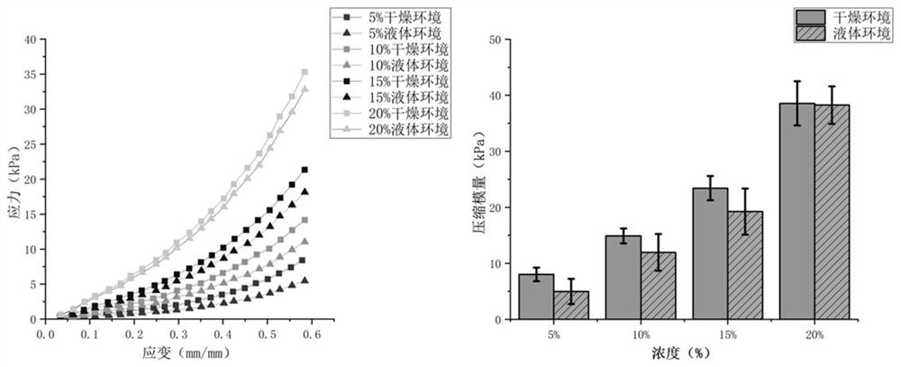
[0080] Example 1 The difference of GelMA concentration in the photosensitive restorative material is formed in a dry environment and a compressive modulus test in a liquid environment

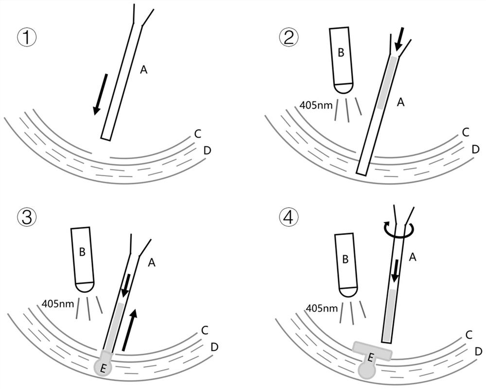
[0081] In the photosensitive repair material prepared by the present invention, the final concentration of methacrylated gelatin GelMA in the photosensitive repair material is 5%-20% (w / v), and the final concentration of polyacrylate in the photosensitive repair material is 5%. -30% (w / v), the final concentration of the photoinitiator in the photosensitive repair material is 0.5%-1% (w / v), and the solvent of the photosensitive repair material is PBS phosphate buffer solution. The inventor found experimentally that the blue light initiator LAP only has an absorption value for light with a wavelength less than 405nm, see Figure 9 , so in the experiments, blue light with a wavelength of 405nm was used for irradiation.

[0082] 1. Configure different concentrations of photosensitive repair materi...

Embodiment 2

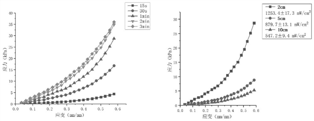
[0099] Example 2 Compression modulus test of the above-mentioned optimal concentration parameters of the photosensitive restoration material prepolymer formed in a liquid environment under different illumination times and different illumination intensities

[0100] 1. Refer to the method of Example 1 to configure the photosensitive restoration material prepolymer whose concentration is the above-mentioned optimal concentration parameter. In the photosensitive repair material, the final concentration of methacrylated gelatin GelMA in the photosensitive repair material is 16% (w / v), and the final concentration of polyacrylate in the photosensitive repair material is 10% (w / v). The final concentration of the agent in the photosensitive repair material is 0.8% (w / v).

[0101] With reference to the method of Example 1, inject this solution into the standard sample mold soaked in Ringer's solution, and immediately irradiate the photosensitive repair material with blue light of 405nm...

Embodiment 3

[0108] Example 3 Effects of Different Polyacrylate Concentrations in Photosensitive Restoration Materials on Tissue Adhesion

[0109] 1. With reference to the photosensitive restoration material formula of Example 1, prepare the photosensitive restoration material prepolymer with polyacrylate concentration gradient, in 4 concentration gradients, the concentration of GelMA is 16% (w / v), LAP photoinitiator The concentration is 0.8% (w / v), the concentration of polyacrylate is 10%, 15% and 20% and 30% respectively, polyacrylate is not added in the control group, other components are consistent with the experimental group, the solvent is PBS phosphate buffer solution.

[0110] 2. Measurement of adhesion characteristic parameters

[0111] Select a glass slide modified with primary amino groups on the surface to simulate the surface of human tissue rich in primary amino groups, drop different concentrations of photosensitive repair materials on the glass slide, and cover one end of ...


PUM

PropertyMeasurementUnit
diameteraaaaaaaaaa

Abstract

The present invention relates to in situ biomanufacturing method in vivo and its application in tissue repair in vivo. The in situ biomanufacturing method in the body provided by the present invention is to transport the photosensitive repair material to the tissue to be repaired in the body through an instrument, fix it as a bioprinting scaffold in the form of a rivet to the tissue to be repaired, and realize the integration of the bioprinting scaffold and the tissue to be repaired by increasing the mechanical force. The good attachment of the repaired tissue solves the problems that the tissue to be repaired is not easy to attach because the tissue to be repaired is located in the deep layer of the body and the surface is wet. The novel photosensitive repair material of the present invention is widely applicable to the in-situ repair of tissues in the body, and the in-situ rapid solidification in the body is riveted with the tissue to be repaired in the form of a hydrogel rivet, and strongly adheres to the tissue. Acrylate is covalently cross-linked with the tissue to be repaired to achieve firm repair and long-term closure of the damaged site, which has a good clinical application prospect.

Description

technical field [0001] The invention relates to the technical field of biomanufacturing and biorepair in vivo, in particular to an in situ biomanufacturing method in vivo and its application in tissue repair in vivo, and also relates to a medical device for in situ tissue in vivo. Background technique [0002] In situ biofabrication refers to the process in which bioinks are directly applied to defective parts of a living body in a clinical setting to create or repair living tissues or organs. The most common form of in situ biomanufacturing is 3D bioprinting. Based on computer-aided additive manufacturing technology, bioink is delivered layer by layer to tissue defects, and the distribution of biomaterials, cells, and growth factors in the three-dimensional structure is precisely controlled. and combined to realize the repair or regeneration of defective tissue. At present, the use of in situ bioprinters for tissue repair has received a lot of attention, and this technolog...

Claims


Application Information

Patent Timeline
Patent Type & Authority Patents(China)
IPC IPC(8): A61L27/16A61L27/22A61L27/50A61F2/00
CPCA61F2/0063A61L27/222A61L27/16A61L27/50A61L2430/34C08L33/08
Inventor 徐弢赵文祥郭晓玲刘正平
Owner TSINGHUA UNIV
Why Eureka
Try Eureka