Application of magnetic exosome to preparation of wound repairing or wound healing product

A technology of wound repair and wound healing, applied in the field of biomedicine, can solve the problems that the safety and curative effect cannot be fully guaranteed, and achieve the effect of great application potential

Inactive Publication Date: 2021-08-20
PEKING UNION MEDICAL COLLEGE HOSPITAL CHINESE ACAD OF MEDICAL SCI
View PDF4 Cites 0 Cited by
  • Summary
  • Abstract
  • Description
  • Claims
  • Application Information

AI Technical Summary

Problems solved by technology

[0005] Although it has been proven that exosomes have good effects in terms of safety and efficacy in treating wounds, their safety and efficacy cannot be fully guaranteed.

Method used


Image

  • Application of magnetic exosome to preparation of wound repairing or wound healing product
  • Application of magnetic exosome to preparation of wound repairing or wound healing product
  • Application of magnetic exosome to preparation of wound repairing or wound healing product

Examples


Embodiment 1

[0039] Example 1: Preparation of Magnetic Exosomes 1

[0040] In this example, magnetic exosomes utilize magnetic Fe 3 o 4 nanoparticles, and magnetic Fe 3 o 4 Magnetic exosomes produced by MSCs stimulated by nanoparticles combined with a static magnetic field.

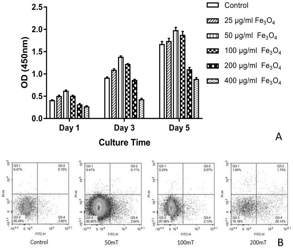
[0041] (1) 10 mg of sterilized Fe 3 o 4 Dissolve in 50ml of complete medium α-MEM, then dilute in equal proportions, respectively configure to concentrations of 400, 200, 100, 50, 25μg / ml containing Fe 3 o 4 culture medium. During the passage of cells, the medium was replaced with different gradients. Determine optimal Fe based on cell growth morphology and proliferation experiments 3 o 4 concentration.

[0042] (2) the best Fe 3 o 4 The cells cultured at different concentrations were stimulated by static magnetic fields (SMF) of different strengths (NdFeB permanent magnet materials, Beijing Xinchangli Functional Materials Co., Ltd.), and the magnetic field strengths were 50, 100, and 150 mT in turn. Det...

Embodiment 2

[0043] Example 2: Cell Proliferation and Apoptosis Experiments

[0044] (1) Cell proliferation experiment: take the MSCs of the P3 generation, digest and centrifuge, resuspend the cells, count, and prepare Fe with a corresponding concentration gradient according to Example 1 3 o 4 -MSC. According to 5×10 3 Inoculate the cell sample into a 96-well plate, add 100 μL of α-MEM medium containing 10% FBS to each well, and culture in an incubator. After 24 hours, discard the medium, add 90 μL of fresh medium and 10 μL of CCK-8 to each well, and incubate at 37° C. for 1 h in the dark. Continuous measurement was carried out for 5 days, with 3 replicate wells per day. The OD value of each well was measured at a wavelength of 450nm, and the proliferation curve was drawn.

[0045] (2) Apoptosis experiment: prepare the Fe with the corresponding magnetic field intensity gradient according to the steps in Example 1 3 o 4 - SMF-MSC, collected directly into a 10ml centrifuge tube after ...

Embodiment 3

[0048] Example 3 Transmission Electron Microscopy and Western Blotting

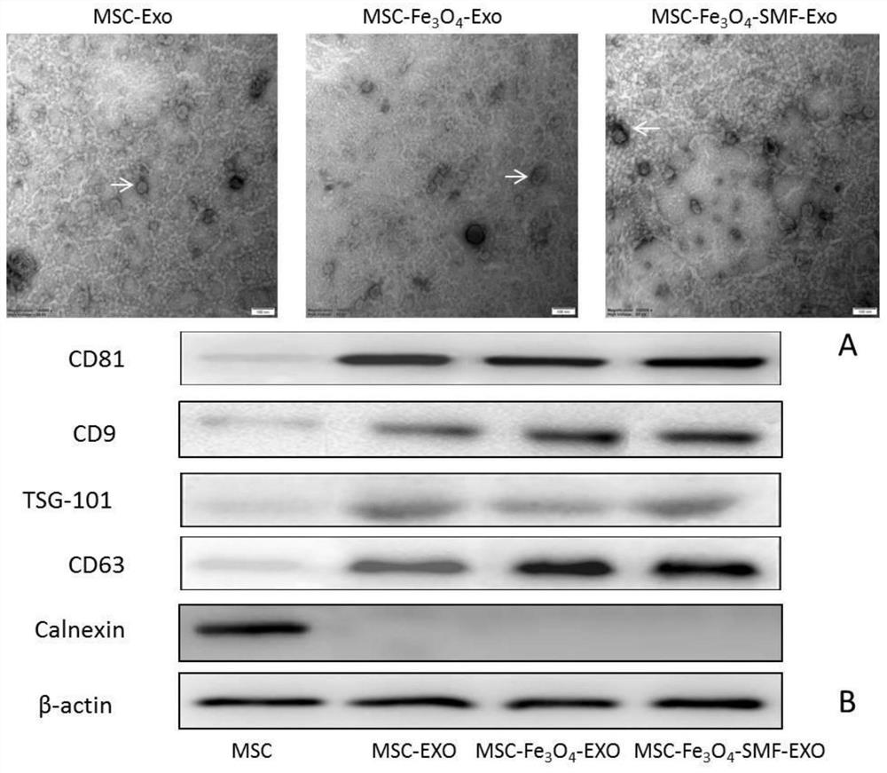
[0049] (1) Observing the morphology of exosomes with the electron microscope uses the optimal concentration of Fe 3 o 4 (50 μg / ml) stimulated exosomes secreted by MSCs were denoted as MSC-Fe 3 o 4 -Exo; optimal concentration Fe 3 o 4 (50 μg / ml) combined with the optimal magnetic field strength (100mT) stimulated the exosomes secreted by MSCs and recorded as MSC-Fe 3 o 4 -SMF-Exo. Take 10 μl of exosomes separated and purified by ultracentrifugation, add an equal volume of balanced salt PBS solution to dilute, and then drop it on a 2 mm sample-loading copper grid. After standing at room temperature for 3 minutes, use filter paper to gently absorb the excess liquid, and use 3% (w / v) 30 μl of sodium phosphotungstate solution (PH6.8) was negatively stained at room temperature for 5 minutes, washed gently with double distilled water, dried at room temperature, observed and photographed under a transmissi...


PUM


Abstract

The invention discloses an application of a magnetic exosome to preparation of a wound repairing or wound healing product. The magnetic exosome is generated by stimulating MSCs (mesenchymal stem cells) by using magnetic Fe3O4 nanoparticles or combining the magnetic Fe3O4 nanoparticles with a static magnetic field. Compared with an untreated mesenchymal stem cell exosome, the magnetic exosome disclosed by the invention has the capability of remarkably enhancing human skin fibroblast proliferation, migration and vascularization. The invention also provides a preparation method of the mesenchymal stem cell exosome. The magnetic exosome prepared by the invention can be used for preparing a preparation for promoting wound repairing and a medicine for treating diabetes wound healing, and has great application potential in tissue regeneration and refractory or huge wound repair or healing treatment.

Description

technical field [0001] The invention relates to the field of biomedical technology, in particular to the application of magnetic exosomes in the preparation of wound repair or wound healing products. Background technique [0002] In many pathological conditions, such as diabetes or severe burns, the normal wound healing process fails to fully restore skin function, leading to potentially serious complications or infection, so it is crucial to complete wound healing after skin tissue damage of. Normal skin tissue is divided into three layers of epidermis, dermis and subcutaneous tissue, and its damage repair is a dynamic and complex process, mainly including: bleeding, coagulation, acute inflammation, cell migration, proliferation and differentiation, angiogenesis, extracellular matrix synthesis and remodeling. These complex processes can basically be summarized into three stages: ①Inflammation stage; ②Proliferation stage of tissue cells; ③Remodeling of tissue structure. ...

Claims


Application Information

Patent Timeline
Patent Type & Authority Applications(China)
IPC IPC(8): A61K35/28A61P17/02A61P3/10
CPCA61K35/28A61P17/02A61P3/10
Inventor 王海吴狄吴志宏邱贵兴
Owner PEKING UNION MEDICAL COLLEGE HOSPITAL CHINESE ACAD OF MEDICAL SCI
Why Eureka
Try Eureka