Glycoprotein N-sugar chain analysis method

An analysis method and glycoprotein technology, applied in the direction of material analysis, material analysis by electromagnetic means, material separation, etc., can solve problems such as the health threat of experimenters, unstable fluorescence signal, increase material cost, etc., to increase popularization, Strong applicability and the effect of increasing detection throughput

Pending Publication Date: 2021-08-24
SHENZHEN TAILI BIOTECHNOLOGY CO LTD
View PDF3 Cites 2 Cited by
  • Summary
  • Abstract
  • Description
  • Claims
  • Application Information

AI Technical Summary

Problems solved by technology

[0006] (1) The pre-processing time for a single sample is too long, and it takes at least 3 working days from getting the sample to getting the result;
[0007] (2) The reducing agent sodium cyanoborohydride used in the marking process of conventional analysis methods is a toxic reagent, which poses a threat to the laboratory environment and the healt

Method used


Image

  • Glycoprotein N-sugar chain analysis method
  • Glycoprotein N-sugar chain analysis method

Examples


Embodiment 1

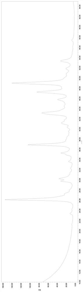
[0066] Take the recombinant human erythropoietin (Fc) fusion protein sample for inspection according to the following steps, and the obtained N-glycan spectrum chromatogram is shown in figure 1 .

[0067] 1) Sample dilution: Dilute the sample to 5 mg / ml with dilution buffer.

[0068] 2) Reduction: Take 15 μl of the diluted sample solution in a 1.5ml EP tube, add 15 μl of 2 mg / ml RapiGest SF solution, mix for 5 seconds, then add 1 μl of 0.5M DTT, mix well and put it in a 37°C incubator Restore for 30min.

[0069]3) Alkylation: after cooling to room temperature, add 2 μl of 0.5M IAM, mix well, and place in the dark at room temperature for 30 min.

[0070] 4) Enzyme digestion: add 4 μl PNGase F (250 U / ml), mix well, and place in a 37° C. incubator for enzyme digestion for 2 hours.

[0071] 5) Drying: Open the cover of the enzyme-digested sample and put it into a vacuum drying oven to slowly evacuate the sample to completely dry the sample.

[0072] 6) Labeling: 5 μl of the pr...

Embodiment 2

[0078] Human IgG samples were taken for testing according to the following steps.

[0079] 1) Sample dilution: Dilute the sample to 5 mg / ml with dilution buffer.

[0080] 2) Reduction: Take 15 μl of the diluted sample solution in a 1.5ml EP tube, add 15 μl of 3mg / ml RapiGest SF solution, mix for 5 seconds, then add 1 μl of 0.5M DTT, mix well and put it in a 34°C incubator Reduction 40min.

[0081] 3) Alkylation: after cooling to room temperature, add 2 μl of 0.5M IAM, mix well, and place in the dark at room temperature for 20 min.

[0082] 4) Enzyme digestion: Add 4 μl PNGase F (300 U / ml), mix well, and place in a 37°C incubator for enzyme digestion for 1.5 hours.

[0083] 5) Drying: Open the cover of the enzyme-digested sample and put it into a vacuum drying oven to slowly evacuate the sample to completely dry the sample.

[0084] 6) Labeling: 5 μl of the prepared labeling reagent was added to the aforementioned enzyme-digested sample, mixed evenly, and placed in a 65° C. ...

Embodiment 3

[0087] Serum transferrin samples were taken for testing according to the following steps.

[0088] 1) Sample dilution: Dilute the sample to 5 mg / ml with dilution buffer.

[0089] 2) Reduction: Take 15 μl of the diluted sample solution in a 1.5ml EP tube, add 15 μl of 1 mg / ml RapiGest SF solution, mix for 5 seconds, then add 1 μl of 0.5M DTT, mix well and put it in a 39°C incubator Restore for 20min.

[0090] 3) Alkylation: After cooling to room temperature, 2 μl of 0.5 M IAM was added, mixed well, and placed at room temperature in the dark for 40 minutes.

[0091] 4) Enzyme digestion: add 4 μl PNGase F (200U / ml), mix well, and place in a 37°C incubator for enzyme digestion for 2.5 hours.

[0092] 5) Drying: Open the cover of the enzyme-digested sample and put it into a vacuum drying oven to slowly evacuate the sample to completely dry the sample.

[0093] 6) Labeling: 5 μl of the prepared labeling reagent was added to the aforementioned enzyme-digested sample, mixed evenly,...


PUM


Abstract

The invention relates to the technical field of biology, in particular to a glycoprotein N-sugar chain analysis method. The method comprises the following steps: a) carrying out protein expansion denaturation and disulfide bond reduction treatment on glycoprotein to be analyzed by using 2-methylpyrimidine borane and a disulfide bond reducing agent; b) carrying out alkylation treatment on the protein treated in the step a), and carrying out enzyme digestion on N-glycosidase F; and c) drying the sample subjected to enzyme digestion, marking the dried sample with a marker, and analyzing the N-carbohydrate chain according to the marker, wherein the marker is procaine amide and/or a salt thereof. The method can obviously shorten the operation process and increase the detection flux, and is more environment-friendly and easy to use.

Description

technical field [0001] The invention relates to the field of biotechnology, in particular to a method for analyzing glycoprotein N-sugar chains. Background technique [0002] During the production of recombinant proteins, conditions such as culture conditions will affect the level of post-translational modification of cellular proteins. Glycosylation is one of the most common post-translational modifications of proteins in eukaryotic cells. It plays a vital role, such as protein folding, cell recognition, cancer cell metastasis, membrane protection, blood group determination system, material transport, etc. [0003] The most common glycoproteins in organisms are N-glycosidic bonded glycoproteins, that is, proteins modified by linking oligosaccharides to the amide nitrogen of the asparagine side chain on the peptide chain. N-glycan chains only account for a small part of the entire glycoprotein, but have a huge impact on the properties of the entire glycoprotein, so N-glycan...

Claims


Application Information

Patent Timeline
IPC IPC(8): G01N30/02G01N30/06G01N30/72G01N1/28G01N27/447
CPCG01N30/02G01N30/06G01N30/72G01N1/28G01N27/447G01N2030/067
Inventor 魏星任兰万军陈亮梁国龙刘友桃
Owner SHENZHEN TAILI BIOTECHNOLOGY CO LTD
Why Eureka
Try Eureka