Fluorescent probe for detecting glucuronyl transferase 1A1 and application thereof

A technology of glucuronic acid and fluorescent probes, which is applied in the field of detecting glucuronyltransferase 1A1 fluorescent probes, can solve the problems of low efficiency, cumbersome detection and poor stability, and achieves the effects of high efficiency, low noise and fast speed.

Active Publication Date: 2021-08-27
DALIAN MEDICAL UNIVERSITY
View PDF6 Cites 0 Cited by
  • Summary
  • Abstract
  • Description
  • Claims
  • Application Information

AI Technical Summary

Problems solved by technology

At present, the detection methods used in clinical practice have problems such as poor stability, cumbersome detection, and low efficiency.

Method used


Image

  • Fluorescent probe for detecting glucuronyl transferase 1A1 and application thereof
  • Fluorescent probe for detecting glucuronyl transferase 1A1 and application thereof
  • Fluorescent probe for detecting glucuronyl transferase 1A1 and application thereof

Examples


Embodiment 1

[0069] Example 1. Confirmation of the structure of the dihydroxy probe

[0070] (1) Determination of the best fluorescent site in the probe

[0071]

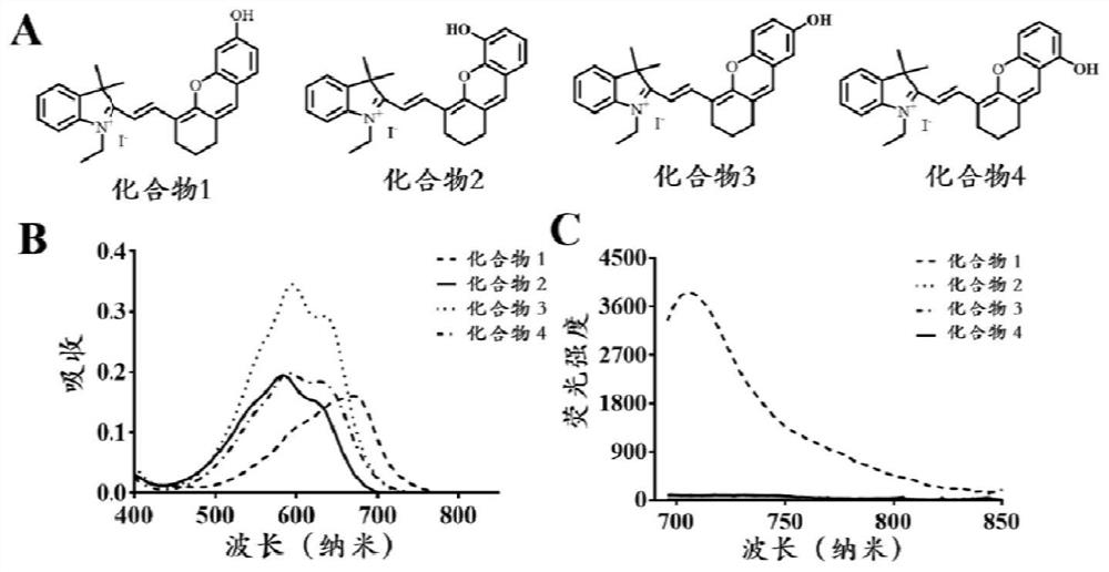
[0072] Hydroxyl-substituted compounds 1-4, wherein the 6-position is substituted by the hydroxy group in compound 1, the 5-position is substituted by the hydroxy group in compound 2, the 7-position is substituted by the hydroxy group in compound 3, and the 8-position is substituted by the hydroxy group in compound 4.

[0073] The absorption spectrum and fluorescence spectrum of compound 1-4 are measured, the absorption spectrum of compound 1-4 in the Tris-HCl system, the collection band is 400-850nm; the fluorescence emission spectrum of compound 1-4 in the Tris-HCl system, the excitation wavelength 670nm, acquisition band 696-850nm. The result is as figure 1 As shown, the maximum absorption of compound 1 is at 690nm, while the maximum absorption of compounds 2-4 is around 580nm; at the same time, the fluorescence spectrum ...

Embodiment 2

[0077] The synthesis of embodiment 2 compound HHC

[0078] The synthetic route of HHC is as follows:

[0079]

[0080] Dissolve 3,4-dimethoxy-2-hydroxybenzaldehyde (1mmol) and 2-bromo-1-cyclohexene-1-carbaldehyde (284mg, 1.5mmol) in 10mL N,N-dimethylformaldehyde To the amide, cesium carbonate (815mg, 2.5mmol) was added and stirred at room temperature for 2h. Then the reaction solution was poured into deionized water, extracted with ethyl acetate, the organic phase was concentrated by vacuum distillation, and the residue was separated by column chromatography (petroleum ether:ethyl acetate=3:1) to obtain a yellow solid. 139mg, yield 51%. 1 H NMR (400MHz, CDCl 3 )δ10.44(s,1H),6.88(d,J=8.5Hz,1H),6.67(d,J=8.5Hz,1H),6.64(s,1H),3.92(s,3H),3.90( s,3H),2.58(dd,J=8.8,3.6Hz,2H),2.46(t,J=6.1Hz,2H),1.76–1.70(m,2H).

[0081] Compound 3 (0.1 mool) and compound 4 (32 mg, 0.1 mmol) were dissolved in 2 mL of acetic anhydride, potassium carbonate (21 mg, 0.15 mmol) was added, and stirre...

Embodiment 3

[0083] Example 3 In vitro determination of the selectivity of HHC to different UGTs single enzymes

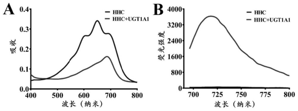
[0084] (1) Prepare 190 μL in vitro metabolic reaction system in advance, including Tris-HCl buffer solution (50mM) at pH 7.4, different kinds of UGT single enzymes (0.1mg / mL), HHC (final concentration 10μM) and shake pre-incubation at 37°C 3 minutes;

[0085] (2) Add 10 μL of UDPGA with a concentration of 40 mM (final concentration 2 mM) to the reaction system to initiate the reaction;

[0086] (3) After 30 minutes, add 100 μL of glacial acetonitrile, shake vigorously, and terminate the reaction;

[0087] (4) Use a high-speed refrigerated centrifuge at 4°C and 20,000×g to centrifuge at high speed for 20 minutes, take the supernatant, and perform fluorescence detection (HHC-G: Ex=670nm, Em=720nm, the results are as follows image 3 shown). Figure 4 It is shown that only UGT1A1 can catalyze the reaction, and the reaction rate is much higher than that of other hydrolases, indi...


PUM

PropertyMeasurementUnit
concentrationaaaaaaaaaa

Abstract

The invention discloses a fluorescent probe for detecting glucuronyl transferase 1A1 and application thereof, and belongs to the technical field of biological medicines. The fluorescent probe can be used for determining the activity of UGT1A1 in biological systems of different sources. HHC is used as a specific probe reaction substrate, by means of a glucuronyl transferase in-vitro reaction system, glucuronylation binding reaction of 5-site hydroxyl of HHC is used as a probe reaction, and the activity of UGT1A1 in each biological sample is determined by quantitatively detecting the generation amount of a metabolite HHC-5-beta-D-Glucuronoside (HHC-G) in unit time. The fluorescent probe can be used for qualitative and quantitative determination of UGT1A1 activity in different individual source human and animal tissue samples, different species of cells and cell preparations, and various plants and microorganisms, can also realize in-vivo imaging research on UGT1A1, and can rapidly evaluate the liver bile excretion function at the animal level. By evaluating the activity of UGT1A1 of different individuals, the method is used for guiding reasonable use of clinical drugs such as irinotecan and high-throughput screening of UGT1A1 inhibitors.

Description

technical field [0001] The invention belongs to the technical field of biomedicine, and in particular relates to a fluorescent probe for detecting glucuronosyltransferase 1A1 (UGT1A1) and an application thereof. Background technique [0002] Glucuronosyltransferase 1A1 (Glucuronosyltransferase 1A1, UGT1A1) is a biphasic detoxification enzyme widely expressed in human liver, small intestine, kidney and other organs. It is mainly distributed in the endoplasmic reticulum, especially in the liver. UGT1A1 can mediate the metabolic detoxification process of a variety of endogenous substances. Most importantly, UGT1A1 is the most important enzyme for the metabolic clearance of human bilirubin, and its activity changes are related to neonatal jaundice and clinical drug-induced jaundice, Hyperbilirubinemia has an inseparable relationship. [0003] The function and expression of UGT1A1 are also easily affected by various factors in the environment, and its activity changes will direc...

Claims


Application Information

Patent Timeline
Patent Type & Authority Applications(China)
IPC IPC(8): C07D405/06C09K11/06G01N21/64A61K31/4745A61K31/366A61P35/00
CPCC07D405/06C09K11/06G01N21/6428A61K31/4745A61K31/366A61P35/00C09K2211/1029C09K2211/1088G01N2021/6439A61K2300/00
Inventor 马骁驰冯磊田象阁崔京南刘涛
Owner DALIAN MEDICAL UNIVERSITY
Why Eureka
Try Eureka