Method for rapidly dyeing biological sample and acquiring three-dimensional data

A biological sample, rapid dyeing technology, applied in the field of biomedical photonics, can solve the problems of limited types of dyes, slow signal acquisition process, incomplete dyeing, etc., to reduce syneresis, accelerate dyeing efficiency, and improve acquisition. The effect of efficiency

Active Publication Date: 2021-08-31
HUST SUZHOU INST FOR BRAINMATICS
View PDF5 Cites 0 Cited by
  • Summary
  • Abstract
  • Description
  • Claims
  • Application Information

AI Technical Summary

Problems solved by technology

[0004] The disadvantage of these methods is that the method of in vivo injection staining is limited to live animals, and due to obstacles such as the blood-brain barrier, the types of optional staining agents are limited, and it will lead to problems such as incomplete staining; if the sample is manually sectioned , staining, and imaging are not only time-consuming and labor-intensive, but also difficult to reflect the real three-dimensional structure of biological tissue samples only for two-dimensional thin section staining and imaging results; the method of soaking and staining isolated tissues for a long time is time-consuming, and generally takes several days for sample staining , which slows down the process of getting the signal
Due to the limited penetration depth of the dye, it is not even possible to achieve whole-body staining for larger volume samples such as rhesus monkey brain

Method used


Image

  • Method for rapidly dyeing biological sample and acquiring three-dimensional data
  • Method for rapidly dyeing biological sample and acquiring three-dimensional data
  • Method for rapidly dyeing biological sample and acquiring three-dimensional data

Examples


Embodiment 1

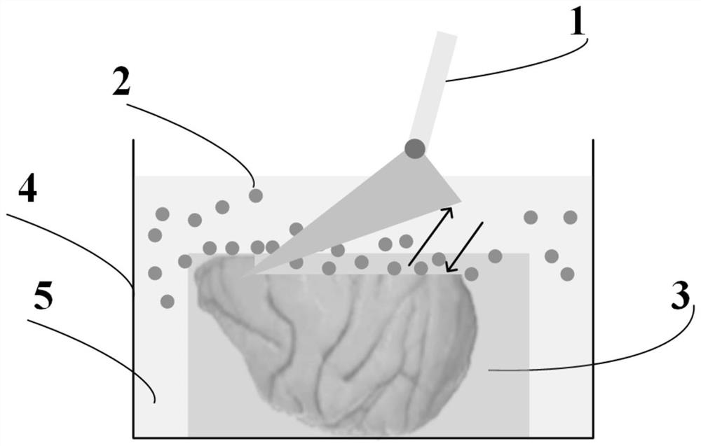
[0068] The imaging solution in Example 1 was composed of an aqueous solution with a concentration of 0.25 wt% Thioflavin S, 30 wt% fructose, 20 wt% urea and 20v / v% DMSO. The biological sample tissue is the brain tissue of 5XFAD transgenic mice, which has the pathological characteristics of β-amyloid plaques, and the three-dimensional data acquisition is completed according to the following steps:

[0069] S1, mice were anesthetized and then perfused. The perfusion solution was firstly 0.01M phosphate buffer solution, and then 4% paraformaldehyde solution, and the brain was taken as the biological sample tissue. The biological sample tissue was soaked in 4% paraformaldehyde solution and fixed for 24 hours until the sample hardened.

[0070] S2. Perform pretreatment on the acquired biological sample tissue, that is, immerse the biological sample tissue in an imaging solution for at least 24 hours. During this process, the imaging solution dehydrates the biological sample tissue...

Embodiment 2

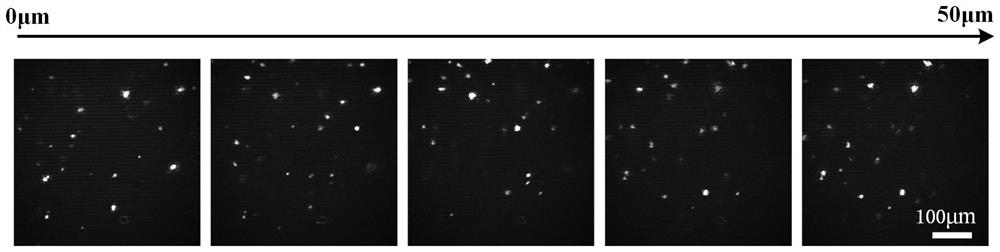
[0082] The imaging solution in Example 2 is composed of 2ug / ml PI (propidium iodide), 30wt% fructose, 20wt% urea and 20v / v% dimethyl sulfoxide aqueous solution. The biological sample tissue is wild-type C57 mouse brain tissue, and the three-dimensional data collection is completed according to the following steps:

[0083] S1, mice were anesthetized and then perfused. The perfusion solution was firstly 0.01M phosphate buffer solution, and then 4% paraformaldehyde solution, and the brain was taken as the biological sample tissue. The biological sample tissue was soaked in 4% paraformaldehyde solution and fixed for 24 hours until the sample hardened.

[0084] S2. Perform pretreatment on the acquired biological sample tissue, that is, immerse the biological sample tissue in an imaging solution for at least 24 hours. During this process, the imaging solution dehydrates the biological sample tissue and makes the surface transparent and stained.

[0085] S3, embedding the pretreat...

Embodiment 3

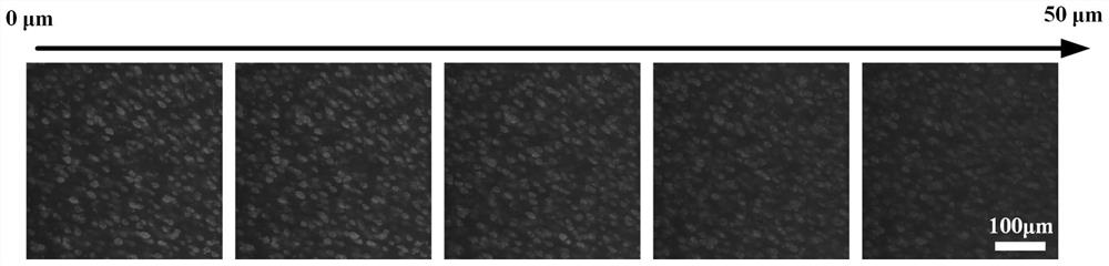
[0096] The imaging solution in Example 3 was composed of DAPI (4',6-diamidino-2-phenylindole), 30wt% fructose, and 20wt% urea at a concentration of 1ug / ml. The biological sample tissue is wild-type C57 mouse brain tissue, and the three-dimensional data collection is completed according to the following steps:

[0097] S1, mice were anesthetized and then perfused. The perfusion solution was firstly 0.01M phosphate buffer solution, and then 4% paraformaldehyde solution, and the brain was taken as the biological sample tissue. The biological sample tissue was soaked in 4% paraformaldehyde solution and fixed for 24 hours until the sample hardened.

[0098] S2. Perform pretreatment on the acquired biological sample tissue, that is, immerse the biological sample tissue in an imaging solution for at least 24 hours. During this process, the imaging solution dehydrates the biological sample tissue and makes the surface transparent and stained.

[0099] S3, embedding the pretreated bi...


PUM


Abstract

The invention discloses a method for rapidly dyeing a biological sample and obtaining three-dimensional data, which comprises the following steps: S1, a first immersion step, S2, an embedding step, S3, a second immersion step, S4, a sample installation step, S5, an optical imaging step, S6, a slicing step; wherein the S5 and S6 are circulated until a three-dimensional image of a biological sample tissue is obtained, and wherein the imaging solution comprises the following raw materials in percentage by mass: 20-40% of sugar or alcohol or sugar alcohol compound; 10-30% of urea; 0-30% of a penetration enhancer aqueous solution; and the balance is the coloring agent. The imaging solution composed of the fructose, the urea, the penetration enhancer and the coloring agent is used for rapidly replacing interstitial fluid in the biological tissue sample, so that the newly exposed surface of the sample is rapidly transparent and dyed; the dyeing efficiency is greatly improved; by matching the microscopic cutting and optical imaging, the three-dimensional data of the dyed biological samples with different volumes can be quickly obtained.

Description

technical field [0001] The invention relates to the technical field of biomedical photonics, in particular to a method for quickly staining biological samples and acquiring three-dimensional data. Background technique [0002] At present, various dyeing techniques are more and more used in biological research, among which fluorescent staining technology is more prominent. Scientists have developed many dyes for specific marking of biological tissues through the research on the interaction between various dyes and biological tissues. People can obtain a series of biological microstructure pictures by combining staining technology with optical microscopy. Due to the development of optical imaging and dyeing and labeling technology, the study of the structure of biological tissue is not limited to thin section, more importantly, it is to obtain the three-dimensional data of the specific structure of biological tissue. [0003] The existing methods of staining and obtaining ima...

Claims


Application Information

Patent Timeline
Patent Type & Authority Applications(China)
IPC IPC(8): G01N1/30G01N21/84
CPCG01N1/30G01N21/84
Inventor 杨孝全张云飞李向宁龚辉
Owner HUST SUZHOU INST FOR BRAINMATICS
Why Eureka
Try Eureka