Placenta protein 13 test strip and quantitative detection method
A quantitative detection method and placental protein technology, which are applied in measurement devices, biological tests, material inspection products, etc., can solve the problems of unsuitable rapid and accurate quantitative detection, high background value, low sensitivity, etc., and achieve reliable diagnostic results and simple operation. , the effect of small size
- Summary
- Abstract
- Description
- Claims
- Application Information
AI Technical Summary
Problems solved by technology
Method used
Image

Examples
Embodiment 1
[0032] A kind of placental protein 13 quantitative detection test strip and detection method of the present invention comprise the following steps:
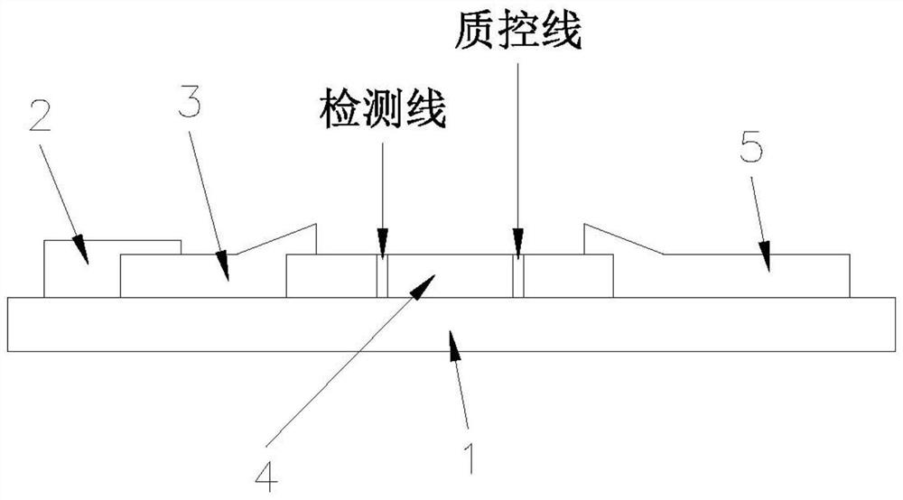
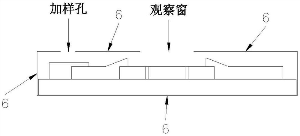
[0033] The test strip comprises a PVC bottom plate 1, and the PVC bottom plate 1 is provided with a sample pad 2, a binding pad 3, a nitrocellulose membrane 4 and a water-absorbing pad 5 in sequence, and the sample pad 2 is lapped on the binding pad 3, and the The bonding pad 3 and the water-absorbing pad 5 are lapped on both ends of the nitrocellulose membrane 4 respectively.
[0034] Quantum dot-labeled anti-placental protein 13 antibody and quantum dot-labeled rabbit anti-bovine serum albumin and dinitrophenol conjugate (rabbit anti-DNP-BSA) are immobilized on the binding pad 3;
[0035] The nitrocellulose membrane 4 is provided with a detection line and a quality control line, the detection line is fixed with an antibody against another epitope of placental protein 13, and the quality control line is fixed with bovine serum a...
Embodiment 2
[0040] In this embodiment, colloidal gold, fluorescent microspheres, gold magnetic nanometers, upconversion luminescence, time-resolved fluorescence, etc. can also be used as tracers. When different tracers are used, the corresponding tracer detection instrument should be changed to an instrument capable of quantitatively detecting the optical intensity of the tracer. Wherein, when the tracer is colloidal gold, the detection instrument is a colloidal gold immunoassay analyzer. When the tracer is a fluorescent microsphere, the detection instrument is a fluorescent immunoassay analyzer. When the tracer is gold magnetic nanometer, the detection instrument is gold magnetic nanometer immune analyzer. When the tracer is an upconversion luminescent material, the detection instrument is an upconversion luminescence immunoassay analyzer. When the tracer is time-resolved fluorescence, the detection instrument is a time-resolved fluorescence analyzer.
[0041] The rest of this embodim...
Embodiment 3
[0043] In this embodiment, quantum dot-labeled goat anti-rabbit IgG is immobilized on the binding pad 3, and rabbit IgG is immobilized on the quality control line;
[0044] The rest of this embodiment is the same as that of Embodiment 1, and the features not explained in this embodiment are explained in Embodiment 1, and will not be repeated here.
PUM
 Abstract
Description
Claims
Application Information