High-resolution spatial omics detection method for tissue sample

A tissue sample and spatial omics technology, which is applied in the field of high-resolution spatial omics detection of tissue samples, can solve the difference of oligonucleotide sequence region modification effect, increase product cost and design complexity, and cross tissue spatial sequence information. Pollution and other problems, to achieve the effect of reducing material costs, reducing instrument costs, and reducing the types of use

Active Publication Date: 2021-11-05
JILIN UNIV
View PDF6 Cites 1 Cited by
  • Summary
  • Abstract
  • Description
  • Claims
  • Application Information

AI Technical Summary

Problems solved by technology

[0006] However, there are still several important issues to be solved for 10×Visium: First, the oligonucleotide sequence array on the surface of the glass slide is prepared by inkjet spotting technology. Each sequence lattice corresponds to the spatial omics information of a dozen to dozens of cells, which limits the analysis of single-cell spatial omics in tissue samples and the exploration of the interaction mechanism between cells, and may also lead to the loss of important information. Omission; Second, when the inkjet spotting method is used to prepare the oligonucleotide sequence array, there will be uneven spotting, which will lead to differences in the modification effect between the oligonucleotide sequence regions, and even the situation of missing spots, that is, No samples were spotted in the target area
Third, when the product is used for sequence capture, the permeabilization fluid promotes the leakage of the sequence in the tissue sample and also causes the sequence to diffuse laterally, resulting in cross-contamination of the sequence information in the tissue space, especially in the high-resolution space group. This phenomenon is particularly serious in the process of scientific sequencing
Fourth, this approach requires separate full-length synthesis of the oligonucleotide capture sequence for each position in the array, and high-volume oligonucleotide synthesis increases the cost and design complexity of the product

Method used


Image

  • High-resolution spatial omics detection method for tissue sample
  • High-resolution spatial omics detection method for tissue sample
  • High-resolution spatial omics detection method for tissue sample

Examples


Embodiment 1

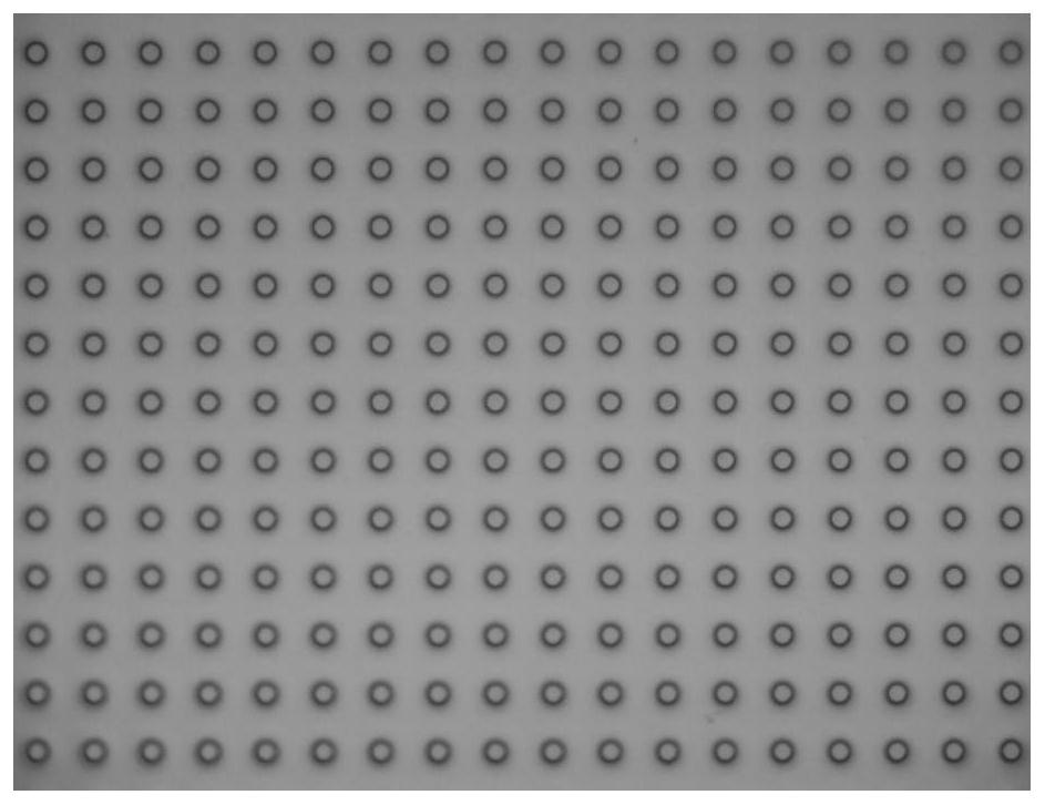
[0138] Embodiment 1: Microwell slide preparation

[0139] Expose the glass plate with a uniform chromium film and photoresist layer under the mask plate of the transparent array for 10s, and then soak the substrate in the developer solution for 30s to obtain a patterned photoresist with a chromium layer. Resist the surface of the array glass slide; then place it in a chromium etching solution and soak it for 5 minutes, then place the glass slide in a glass etching solution (mass ratio HF:HNO 3 : NH 4 F: H 2 (0=25:23.5:9.35:450) soaked in 80min to obtain the patterned microwell array glass slide ( figure 1 ), where the height of the microwells is 40 μm, the diameter is 30 μm, and the distance between the centers of the microwells is 60 μm.

Embodiment 2

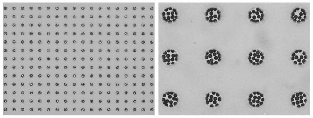
[0140] Embodiment 2: preparation of micro reaction chamber

[0141] Spread the microbead carrier suspension with a diameter of 5-7 μm on the surface of the microwell slide, let it stand for 5 minutes, the microbeads settle in the microwell array, wash the surface of the microwell slide, and wash away the microbeads outside the microwell array, A microwell reaction chamber array containing microbeads was obtained ( figure 2 ).

Embodiment 3

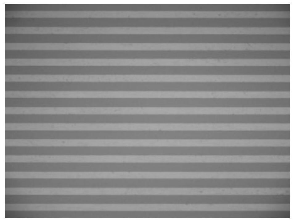
[0142] Embodiment 3: the preparation of microchip

[0143] Place the glass plate with a uniform chrome film and photoresist layer under a mask with parallel holes and expose it to a UV lamp for 10s, then soak the substrate in a developer solution for 30s to obtain micropores with a chromium layer patterned photoresist glass surface; then place it in chrome etching solution for 5 minutes, then place the surface in glass etching solution (mass ratio HF:HNO 3 : NH 4 F: H 2 O=25:23.5:9.35:450) soaked in 40min to obtain the glass channel mold of the morphology structure of the microchannel pattern; polydimethylsiloxane (PDMS) prepolymer and curing agent are in mass ratio of 10:1 Mix the ratio evenly, and after vacuum degassing for 30 minutes, pour it onto the surface of the glass mold, place it in an oven with a temperature of 60°C, cure it for 10 hours, and lift it off to obtain PDMS microfluidic channels arranged in parallel ( image 3 ), the channel height is 20 μm, the width...


PUM

PropertyMeasurementUnit
pore sizeaaaaaaaaaa
widthaaaaaaaaaa
heightaaaaaaaaaa

Abstract

The invention is applicable to a high-resolution spatial omics detection device, system and method for a tissue sample, and provides the high-resolution spatial omics detection device, system and method for the tissue sample, respectively comprising a slide with a micro-well reaction chamber array capable of accommodating a micro-carrier, a method for modifying nucleic acid molecule identifiers and a method for reducing cross contamination of omics information in the process of capturing spatial omics information of the tissue sample. By adopting the spatial omics detection method disclosed by the invention, the resolution ratio of spatial omics detection is remarkably improved, the detection cost is reduced, and meanwhile, the cross contamination of spatial omics information is fundamentally reduced.

Description

technical field [0001] The present invention mainly relates to a high-resolution spatial omics detection device, system and method for tissue samples, in particular to a high-resolution spatial omics detection method for tissue samples. Background technique [0002] Human tissue is a highly complex system composed of trillions of cells, which vary in type, time and space. For example, tissues in different regions of the mammalian brain have different functions and cell types. Qualitative testing is particularly important. Spatial omics refers to the omics research that is done on tissue slices and retains the spatial information of the sample. Spatial omics can display the gene expression in different regions of tissue slices, reveal the activated signaling pathways in fine pathological regions, and complete the mechanism analysis of molecular features driving pathological features. Space omics has completed the technical innovation of pathological digitalization combined ...

Claims


Application Information

Patent Timeline
Patent Type & Authority Applications(China)
IPC IPC(8): C12Q1/6874
CPCC12Q1/6874C12Q2563/159C12Q2563/143C12Q2563/149C12Q2521/107C12Q2531/113C12Q2565/519Y02A90/10
Inventor 张俊虎梁重阳于年祚金正洋杨柏
Owner JILIN UNIV
Why Eureka
Try Eureka