Method for determining minimal residual lesion level based on multiple amplification sequencing

A micro-residue, multiple amplification technology, applied in biochemical equipment and methods, microbial determination/inspection, resistance to vector-borne diseases, etc., can solve the problem of ineffective removal of PCR preference, inaccurate and effective MRD level detection Problems such as low data ratio, to avoid the generation of primer-dimers, reduce UMI overlap, and improve efficiency

Active Publication Date: 2022-05-13
QIAGEN SUZHOU TRANSLATIONAL MEDICINE CO LTD
View PDF9 Cites 0 Cited by
  • Summary
  • Abstract
  • Description
  • Claims
  • Application Information

AI Technical Summary

Problems solved by technology

In the prior art, the traditional two-step method of amplicon library construction and sequencing is used to detect the BCR rearrangement to evaluate the MRD level, which is not only relatively cumbersome to operate, but also needs to be quantified by

Method used


Image

  • Method for determining minimal residual lesion level based on multiple amplification sequencing
  • Method for determining minimal residual lesion level based on multiple amplification sequencing
  • Method for determining minimal residual lesion level based on multiple amplification sequencing

Examples


Embodiment 1

[0093] Example 1: Plasmid DNA Primer Amplification Test

[0094] Design and verification of target binding primers: download the reference sequences of immunoglobulin IgH, IgK, and IgL from public databases, and screen the homologous conserved regions through sequence comparison analysis for primer design. A total of 25 specific primers were designed. SEQ ID NO .1~25:

[0095] SEQ ID NO. 1 (IgHV1): ACAGYCTACATGGAGCTGAG

[0096] SEQ ID NO. 2 (IgHV2): GACCAACATGGACCCTGTGGA

[0097] SEQ ID NO. 3 (IgHV3): GGGCCGRTTCACCATCTCCA

[0098] SEQ ID NO. 4 (IgHV4): ACAGCCTGAAAACCGAGGACA

[0099] SEQ ID NO. 5 (IgHV5): CATCTCCAGAGACAATTCCARGAAC

[0100] SEQ ID NO. 6 (IgHV6): GCTGAACTCTGTGACTCCCGAGG

[0101] SEQ ID NO. 7 (IgHV7): GACACCTCTGCCAGCACAGCAT

[0102] SEQ ID NO. 8 (IgHJ): CTCACCTGAGGAGACAGTGACC

[0103] SEQ ID NO.9 (IgKV1): GGTTCAGYGGCAGTGGATCT

[0104] SEQ ID NO. 10 (IgKV2): ACWGATTTYACACTGAAAATCAGC

[0105] SEQ ID NO. 11 (IgKV3): CAGGCTCCTCATCTATGRTGCATC

[0106] SEQ ID NO...

Embodiment 2

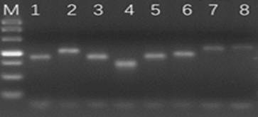
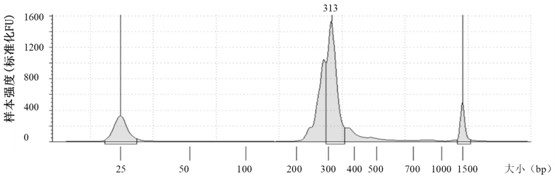
[0129] Example 2: Cell Line DNA Studies

[0130] In this embodiment, the verification of MRD level detection is firstly performed on cell lines known in the public database. Specific steps are as follows:

[0131] 1. Screen cell lines:

[0132]

[0133] 2. Cell line nucleic acid preparation: All 6 cell lines were purchased from ATCC. Take 1mL of cell suspension and use the whole blood DNA extraction kit of Meijie to extract nucleic acid according to the instructions. The extracted nucleic acid was concentrated using Qubit3.0 and Agilent2100 and purity determination, and stored at -20°C for future use.

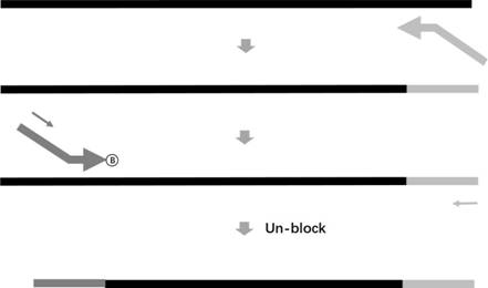
[0134] 3. Make the primer pool: refer to the primer sequence of SEQ ID NO.26~50, mix the 25 synthesized primers according to a certain ratio to obtain the forward primer pool and the reverse primer pool, of which SEQ ID NO.26~46 It is a forward primer (with a blocking modification group C3 at the 3' end), and SEQ ID NO.47~50 is a reverse primer. The number of forward prim...

Embodiment 3

[0212] Embodiment 3: clinical sample research

[0213] It has been verified in Example 2 that the detection result of the method for measuring MRD level of the present invention is accurate and effective for known cell lines, and this example uses clinical samples as materials for further verification. Specific steps are as follows:

[0214] 1. Clinical sample collection:

[0215] Samples of 15 patients with hematological malignancies were collected from cooperative hospitals. They may suffer from different diseases. The sample types include bone marrow fluid and peripheral blood. The patients were between 3 and 74 years old. There were 8 males and 7 females, see Table 2.

[0216] Table 2

[0217]

[0218] 2. Sample processing:

[0219]All 15 samples (bone marrow fluid and peripheral blood) were extracted using the Maijie Whole Blood DNA Extraction Kit according to the operating instructions.

[0220] 3. Amplified sequencing analysis: the steps are the same as in Exampl...


PUM


Abstract

The invention discloses a method for determining the minimal residual lesion level based on multiple amplification sequencing. Comprising the following steps: S1, sample treatment, S2, primer pool manufacturing, S3, single-ended amplification, S4, quality control, S5, purification, S6, mixed sequencing, S7, sequencing data analysis, and S8, minimal residual lesion level calculation. According to the determination method, simple and convenient single-ended amplification is used, the minimal residual lesion level can be accurately and quantitatively detected, an external reference is not needed, meanwhile, primer dimers can be prevented from being generated, the proportion of effective data is increased, and then the accuracy of determining the MRD level is improved.

Description

technical field [0001] The invention relates to the field of biotechnology, in particular to a method for determining the level of minimal residual disease based on multiple amplification sequencing. Background technique [0002] B cell antigen receptor (BCR) is an immunoglobulin molecule (Ig) grown on the surface of B lymphocytes, which contains two heavy chains (H) and two light chains (L). The heavy chain consists of a variable region (VH) and three constant regions (CH1 / CH2 / CH3), while the light chain consists of a variable region (VL) and a constant region (CL). In the variable region, there are four framework regions (FR1 / FR2 / FR3 / FR4) with relatively small changes and three complementarity-determining regions (CDR1 / CDR2 / CDR3) with relatively large changes, of which the largest change is CDR3 Area. According to the gene segment encoding the variable region, the heavy chain can be divided into V-D-J regions, and the light chain can be divided into V-J regions. [0003...

Claims


Application Information

Patent Timeline
IPC IPC(8): C12Q1/6886C12Q1/6844C12Q1/6869C12Q1/6806C40B50/06
CPCC12Q1/6886C12Q1/6844C12Q1/6869C12Q1/6806C40B50/06C12Q2537/143Y02A50/30
Inventor 林东旭刘佳王霞尹松松顾凯丽张亚飞
Owner QIAGEN SUZHOU TRANSLATIONAL MEDICINE CO LTD
Why Eureka
Try Eureka