Tissue engineering liver based on plant acellular scaffold and preparation method

A decellularized scaffold and tissue engineering technology, which is applied in the field of tissue engineering liver and preparation based on plant decellularized scaffolds, can solve the problems of limited usability, achieve suitable porosity and hydrophilicity, high biocompatibility, and simple method Effect

Active Publication Date: 2022-08-02
NANJING DRUM TOWER HOSPITAL
View PDF5 Cites 0 Cited by
  • Summary
  • Abstract
  • Description
  • Claims
  • Application Information

AI Technical Summary

Problems solved by technology

[0002]In recent years, liver decellularized scaffolds refer to the removal of immunogenic cell components while retaining non-immunogenic ECM, retaining the natural 3D structure of the tissue, and providing the next step The cell-to-cell interaction and cell-ECM interaction provide biomaterials with a stable physical structure spac

Method used


Image

  • Tissue engineering liver based on plant acellular scaffold and preparation method
  • Tissue engineering liver based on plant acellular scaffold and preparation method
  • Tissue engineering liver based on plant acellular scaffold and preparation method

Examples


Embodiment 1

[0023] Example 1 Preparation of tissue engineered liver based on plant decellularized scaffolds

[0024] A tissue-engineered liver on a plant decellularized scaffold is prepared by the following method, and the specific steps are as follows:

[0025] S1, intercept the stem of cress with a hollow hole-like structure and clean it;

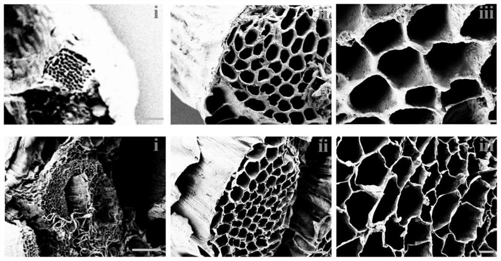
[0026] S2. Submerge the celery stems in 10wt% sodium dodecyl sulfate (SDS) solution for 5 days, and change the solvent every day; The stems were rinsed for two days to obtain decellularized scaffolds, and it could be observed that the fresh celery tissue changed from green to transparent (the tissue cell components were removed); the obtained decellularized scaffolds were washed with PBS solution, and the residual solvent components were used to provide suitable the environment in which cells live; figure 1 The porous structure of the natural celery stem and the prepared decellularized scaffold was shown;

[0027] S3. Mix the hepatocyte mixture wi...

Embodiment 2

[0028] Example 2 Test of compatibility of tissue engineering liver based on plant decellularized scaffolds

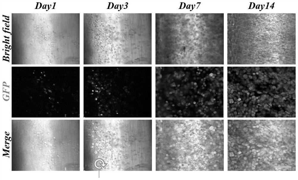
[0029] In order to examine the biocompatibility of the decellularized scaffolds, the tissue engineered liver prepared in Example 1 was cultured for another 14 days, and the hepatocytes suspended in Matrigel were seeded on a 24-well plate as a control group. like figure 2 Cell numbers were determined using GFP staining as indicated. On the third day after seeding, the number of GFP-positive cells was higher than on the first day. In addition, the number of GFP-positive cells increased with the culture time, indicating that the decellularized scaffolds promoted cell proliferation, confirming their high biocompatibility.

Embodiment 3

[0030] Example 3 Characterization of liver function of tissue engineered liver based on plant decellularized scaffolds

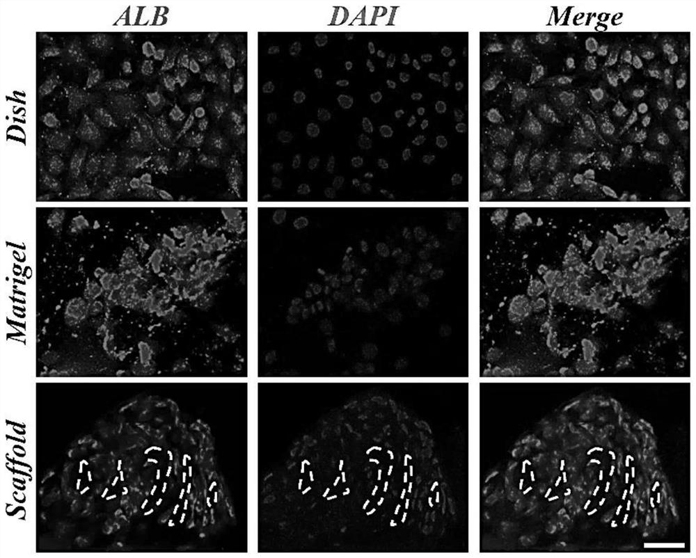
[0031] Due to the lack of extracellular matrix components and cell-to-cell interactions, hepatocytes lose their differentiated state over time when cultured in 2D dishes. However, the composition of Matrigel is heterogeneous, with more than 1500 different proteins in its composition, including the most common proteins such as laminin and type IV collagen. To assess the metabolic activity of transplanted hepatocytes in decellularized liver scaffolds, the albumin secretion, glycogen synthesis, and mRNA expression levels of key hepatic transcription factors and functional genes were quantitatively analyzed. like image 3 As shown, fluorescence images of albumin confirmed that the number of albumin-positive cells was greater in both the Matrigel and decellularized scaffold groups compared to the dish group. Hepatocyte-specific gene expression was performed dur...


PUM


Abstract

The invention provides a preparation method of a tissue engineering liver based on a plant decellularized scaffold. The preparation method comprises the following steps: S1, cutting out a stem part of hollow oenanthe javanica; s2, immersing the stem part of the celery into a lauryl sodium sulfate solution, and then rinsing the stem part of the celery by utilizing trition x-100 containing hypochlorous acid, so as to obtain a decellularized scaffold; and S3, planting the hepatocytes on the acellular scaffold and culturing the hepatocytes.

Description

technical field [0001] The invention belongs to the field of biomedical materials, in particular to a tissue engineering liver based on a plant decellularized scaffold and a preparation method. Background technique [0002] In recent years, liver decellularized scaffolds refer to the removal of immunogenic cellular components while retaining non-immunogenic ECM, preserving the natural 3D structure of the tissue, and providing a stable physical structure for subsequent cell-to-cell interactions and cell-ECM interactions. Biomaterials in different spaces and microenvironments, which promote the formation of new tissues and other characteristics play an important role in tissue engineering and have been widely used. Commonly used liver decellularized scaffolds are mainly derived from human and animal livers. Due to species differences, this May lead to inevitable immune rejection and thus limited availability. Therefore, in the field of biomedicine, there is an urgent need for...

Claims


Application Information

Patent Timeline
IPC IPC(8): C12N5/071A61L27/36A61L27/38A61L27/50A61L27/56
CPCC12N5/0671A61L27/3637A61L27/3641A61L27/3687A61L27/3804A61L27/3839A61L27/50A61L27/56C12N2513/00C12N2533/90A61L2430/40A61L2430/28A61L2300/412
Inventor 任昊桢王经琳赵远锦
Owner NANJING DRUM TOWER HOSPITAL
Why Eureka
Try Eureka