Tissue vascularization promoting gene-chitosan nanometer granula complex

A technology of chitosan nanoparticles and complexes, applied in tissue culture, genetic engineering, plant gene improvement, etc., to achieve the effects of improving vascularization level, large specific surface area, and high expression

Inactive Publication Date: 2005-01-19
INST OF BASIC MEDICAL SCI ACAD OF MILITARY MEDICAL SCI OF PLA
View PDF0 Cites 4 Cited by
  • Summary
  • Abstract
  • Description
  • Claims
  • Application Information

AI Technical Summary

Problems solved by technology

However, chitosan nanoparticles-pcDNA 3.1 -hVEGF recombinant gene is reloaded with type I collagen to form a tissue engineering scaffold, inoculated with cardiomyocytes, cultured in vitro, transplanted into the body, and continued to be cultured in vivo to promote myocardial vascularization, but there is no report

Method used


Image

  • Tissue vascularization promoting gene-chitosan nanometer granula complex

Examples


Embodiment 1

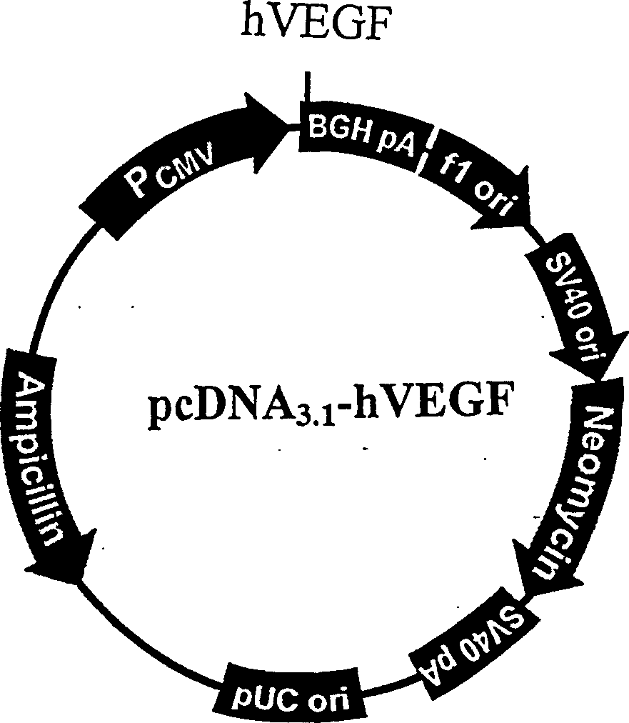
[0010] Embodiment 1 Construction of recombinant pcDNA3.1-hVEGF plasmid

[0011] Plasmid pcDNA3.1 is a eukaryotic expression vector, which constructs a human cytomegalovirus promoter, which can express and regulate the inserted foreign gene. The plasmid itself carries the gene coding sequence of hVEGF, and can express and produce hVEGF under the action of its own expression control elements. There are 17 restriction restriction sites on the multiple cloning site of the plasmid. The BamHI and KpnI regions can be cut with restriction endonucleases, and then T4DNA ligase is used to connect the exogenous gene that has also been double-digested with BamHI and KpnI, so that it is placed under the control of the CMV promoter of human cytomegalovirus, In this way, the eukaryotic expression vector of the foreign gene is constructed (see accompanying drawing).

Embodiment 2

[0012] The structure of embodiment 2 gene-chitosan nanoparticle complex

[0013] It is prepared by coacervation method, and all operations are carried out under sterile conditions. First prepare a 0.03% chitosan (Sigma) solution, adjust it to pH 5.5 with 1N HCl, sterilize with a 0.22um filter membrane, and use a 75 μM sodium sulfate (analytical grade, Beijing Chemical Plant) solution (filtered with a 0.22um filter membrane) Sterilization) dissolve the pcDNA3.1-hVEGF recombinant plasmid to make its final concentration 200ug / ml. Take 200ul of 0.03% chitosan solution and 200ul of 75mM Na containing the recombinant plasmid pcDNA3.1-hVEGF 2 SO 4 The solutions were placed in 55°C water for 20 minutes at a constant temperature. Accurately absorb 200ul Na 2 SO 4 The solution was added to the chitosan solution, and quickly mixed on a vortex mixer for 30 seconds to prepare a suspension of the gene-chitosan nanoparticle complex.

Embodiment 3

[0014] Example 3 Construction of tissue engineered myocardium

[0015] Preparation of type I collagen scaffold: take a rat with a weight of about 250 grams, cut off the tail from the base of the tail, soak it in 75% alcohol for 30 minutes; cut the tail into 1.5cm small pieces under sterile conditions, remove the fur, and draw out Put the tail tendon on a plate, immerse in 150ml of acetic acid solution (0.01-0.25M), sterilize 10 pounds for 10 minutes under high pressure, put it in a refrigerator at 4°C, transfer it to a sterilized centrifuge tube after 48 hours, and centrifuge at 4000 rpm for 30 minutes. Draw the supernatant into vials and store in -20°C refrigerator for later use

[0016] Construction of tissue-engineered myocardium: Type I collagen was used as a scaffold to inoculate cardiomyocytes from suckling rats (1-2 days old). The preparation method of the cardiomyocytes is to take the myocardial tissue of suckling rats, chop and grind them into a uniform cell suspensi...


PUM

PropertyMeasurementUnit
particle sizeaaaaaaaaaa
particle diameteraaaaaaaaaa

Abstract

The invention relates to a tissue vascularization promoting gene-chitosan nanometer particle complex, and belongs to tissue engineering field. The invention inoculates cardiac cell by type 1 collagen of pcDNA#-[3.1]-hVEGF gene-chitosan nanometer particle complex, then transplants them to injured cardiac tissue after in vitro cultivation thereof. Slow releases in vivo and transfects peripheral cardiac cell to obtain target gene expression. Expressed product hVEGF can promote the regeneration of vascular tissue. The growth promoting function of cardiac vessel is intense since the nanometer carry high quantity of gene due to its small volume and specific surface area.

Description

technical field [0001] The invention relates to a gene-chitosan nanoparticle complex for promoting tissue vascularization, which belongs to the field of tissue engineering. Background technique [0002] Due to the particularity of its structure, nanomaterials exhibit many physical and chemical properties different from traditional materials. As a basic constituent unit, nanomaterials are arranged in three dimensions. At present, in the medical field, nanomaterials have been successfully applied. The most notable ones are used as drug carriers, gene carriers or to make human biomedical materials, such as artificial kidneys, artificial joints, etc. In the study of nanomaterials, the current hot spot is nanoparticle gene transfer carrier and drug nanocarrier technology. This technology uses nanoparticles as the carrier of gene transfer. Gene molecules such as DNA and RNA are wrapped in nanoparticles or adsorbed on their surfaces. At the same time, the surface of the particles...

Claims


Application Information

Patent Timeline
Patent Type & Authority Applications(China)
IPC IPC(8): A61L31/12A61L31/16C12N5/077C12N15/12C12N15/79C12N15/85
Inventor 王常勇齐子荣郭希民段翠密赵云山
Owner INST OF BASIC MEDICAL SCI ACAD OF MILITARY MEDICAL SCI OF PLA
Why Eureka
Try Eureka