Eureka AIR delivers breakthrough ideas for toughest innovation challenges, trusted by R&D personnel around the world.

Mutations that confer genetic stability to additional genes in influenza viruses

a technology of influenza viruses and mutations, applied in the field of respiratory pathogens, can solve the problems of significantly attenuated viruses, inaccurate reflection of natural, and insufficient understanding of influenza virus-induced pathology, and achieve the effect of enhancing the stability of influenza viruses and increasing genetic stability

Active Publication Date: 2018-08-21
WISCONSIN ALUMNI RES FOUND
View PDF178 Cites 10 Cited by
  • Summary
  • Abstract
  • Description
  • Claims
  • Application Information

AI Technical Summary

Benefits of technology

[0006]The present invention relates to mutations in influenza virus gene segment(s) that increase genetic stability, for instance, of an additional, non-influenza viral gene, such as a “heterologous” gene sequence that is inserted into one of the viral gene segments, e.g., fused to an intact or modified (for example, truncated or internally deleted) viral protein coding region, or that is present on an additional gene segment. In one embodiment, one or more of the mutations may be employed to enhance the stability of influenza viruses that are not augmented with heterologous gene sequences. In one embodiment, the heterologous gene sequence is a marker gene, e.g., a fluorescent protein gene such as one for GFP, BFP, RFP, or YFP, a luciferase gene, a beta-glucuronidase gene, or beta-lactamase gene. In one embodiment, the heterologous sequence is for a prophylactic gene product. In one embodiment, the heterologous sequence encodes a therapeutic gene product.
[0007]As disclosed herein, influenza viruses expressing fluorescent proteins of different colors (Color-flu viruses) were generated. Viruses containing the foreign matter gene were passaged. Upon adaptation to mice, stable expression of the fluorescent proteins in infected animals allowed their detection by different types of microscopy and by flow cytometry. The use of fluorescent influenza viruses, each of which stably expresses one of four different fluorescent proteins, allows for simultaneous monitoring and live imaging. Using these viruses, several studies were performed to demonstrate the versatility of these viruses. For example, this system was used to analyze the progression of viral spread in mouse lungs, for live imaging of virus-infected cells, and for differential gene expression studies in virus antigen-positive and -negative live cells in the lungs of Color-flu-infected mice. Thus, Color-flu viruses are powerful tools to analyze virus infections at the cellular level in vivo to better understand influenza pathogenesis. Moreover, different stabilizing mutations in the resulting viruses were identified. These mutations include the T380A in HA protein (numbering is that for H1) and E712D in PB2 protein of A / PR / 8 / 34 (H1N1) virus, and V25A, R443K, K737R and P167S amino acid replacements in the PB2, PA, PB1 and NS1 proteins of A / Vietnam / 1203 / 2004 (H5N1) virus, respectively. The individual mutations in the H5 virus alone resulted in the virus containing a foreign gene more stable in vitro, and the combination of all of them provided even greater stability in vivo. These mutations are useful for any HA / NA combination.

Problems solved by technology

Although several studies have addressed host responses to influenza virus infections (Fakuyama and Kawaoka, 2011), the mechanisms of influenza virus-induced pathology are still not fully understood.
However, these viruses were significantly attenuated (Kittel et al., 2004; Shinya et al., 2004) and may not accurately reflect natural infections.
However, the GFP gene was not stably maintained during replication in mouse lung or cultured cells (Manicassamy et al., 2010).
Highly pathogenic avian influenza viruses (HPAI) of the H5N1 subtype continue to evolve in nature, threatening animal and public health.

Method used


Image

  • Mutations that confer genetic stability to additional genes in influenza viruses
  • Mutations that confer genetic stability to additional genes in influenza viruses
  • Mutations that confer genetic stability to additional genes in influenza viruses

Examples


example i

Method

Generation of Color-Flu

[0110]The NS segments of PR8 fused with different fluorescent reporter genes including eCFP, eGFP, Venus, and mCherry were constructed by overlapping fusion PCR as described in Manicassamy et al. (2010). In brief, the open reading frame (ORF) of the NS1 gene without the stop codon was fused with the N-terminus of fluorescent reporter genes via a sequence encoding the amino acid linker GSGG. The fluorescent reporter ORFs were followed by a sequence encoding the GSG linker, a foot-and-mouth virus protease 2A autoproteolytic site with 57 nucleotides from porcine teschovirus-1 in Manicassamy et al. (2010), and by the ORF of nuclear export protein (NEP) (FIG. 5). In addition, silent mutations were introduced into the endogenous splice acceptor site of the NS1 gene to abrogate splicing (Basler et al., 2001). The constructed NS segments (designated eCFP-NS, eGFP-NS, Venus-NS, and mCherry-NS) were subsequently cloned into a pPoll vector for reverse genetics as d...

example ii

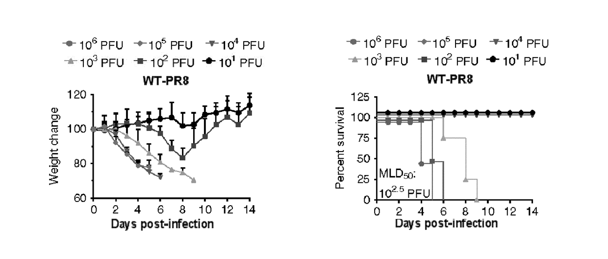
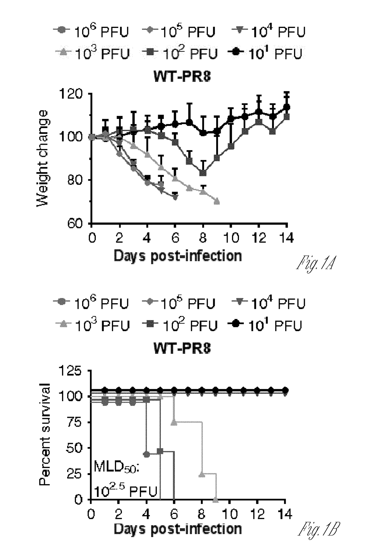
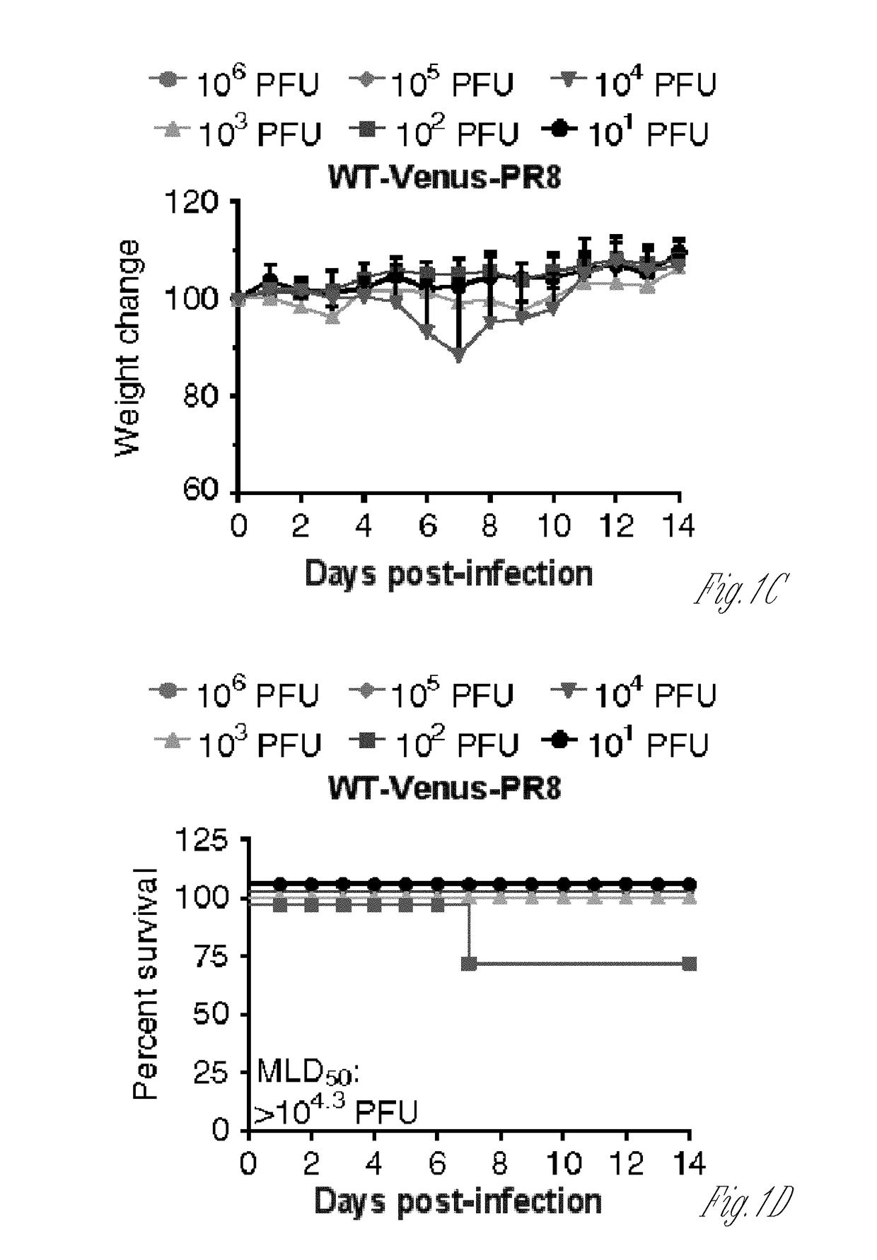
[0133]As disclosed in Example I, an H5N1 virus with the Venus (Nagai et al., 2002) (a variant of eGFP; ref) reporter gene (designated wild-type Venus-H5N1 virus, and abbreviated as WT-Venus-H5N1 virus) was prepared using reverse genetics; this virus showed moderate virulence and low Venus expression in mice. After six passages in mice a mouse-adapted Venus-H5N1 virus was acquired (abbreviated as MA-Venus-H5N1 virus) that stably expressed high levels of Venus in vivo and was lethal to mice; a dose required to kill 50% of infected mice (MLD50) was 3.2 plaque-forming units (PFU), while that of its parent WT-Venus-H5N1 virus was 103 PFU. However, the mechanism for this difference in virulence and Venus stability was unclear.

[0134]In this study, the molecular mechanism that determines the virulence and Venus stability of Venus-H5N1 virus in mice was explored. By using reverse genetics, various reassortants between WT-Venus-H5N1 and MA-Venus-H5N1 virus were rescued and their virulence in ...

example iii

Materials and Methods

[0178]Cells and Viruses.

[0179]Madin-Darby canine kidney (MDCK) cells were maintained in minimum essential medium (MEM) containing 5% of newborn calf serum (NCS). Human embryonic kidney 293T (HEK293T) and HEK293 cells were maintained in Dulbecco's modified Eagle medium supplemented with 10% fetal calf serum (FCS). A / Puerto Rico / 8 / 34 (H1N1; PR8) (Horimoto et al., 2007) and each NS1-Venus PR8 virus were generated by using reverse genetics and were propagated in MDCK cells at 37° C. for 48 hours in MEM containing L-(tosylamido-2-phenyl) ethyl chloromethyl ketone (TPCK)-treated trypsin (0.8 μg / mL) and 0.3% bovine serum albumin (BSA) (Sigma Aldrich).

[0180]Adaptation of NS1-Venus PR8 Virus in Mice.

[0181]Six- to eight-week-old female C57BL / 6 mice (Japan SLC) were intranasally infected with 50 μL of 2.3×10⁶ plaque-forming units (PFU) of NS1-Venus PR8 virus. Lungs were harvested 3-6 days post-infection (dpi) and homogenized in 1 mL of phosphate-buffered saline (PBS). To o...


PUM

PropertyMeasurementUnit
volumeaaaaaaaaaa
wavelengthaaaaaaaaaa
total volumeaaaaaaaaaa

Abstract

The disclosure provides for an isolated recombinant influenza virus having at least one of: a PA gene segment encoding PA with a residue at position 443 that is not arginine, a PB1 gene segment encoding PB1 with a residue at position 737 that is not lysine, a PB2 gene segment encoding PB2 with a residue at position 25 that is not valine or a residue at position 712 that is not glutamic acid, a NS gene segment encoding a NS1 with a residue at position 167 that is not proline, a HA gene segment encoding a HA with a residue at position 380 that is not threonine, or any combination thereof, and methods of making and using the virus.

Description

CROSS-REFERENCE TO RELATED APPLICATIONS[0001]This application claims the benefit of the filing date of U.S. application Ser. No. 62 / 015,074, filed on Jun. 20, 2014, the disclosure of which is incorporated by reference herein.STATEMENT OF GOVERNMENT RIGHTS[0002]This invention was made with government support under HHSN266200700010C awarded by the National Institutes of Health. The government has certain rights in the invention.BACKGROUND[0003]Influenza A virus is a respiratory pathogen that causes annual epidemics and sporadic pandemics (Wright et al., 2013). Moreover, highly pathogenic avian H5N1 and the recently emerged H7N9 influenza viruses have caused an appreciable number of human infections with high mortality rates (Watanabe et al., 2013; Zhang et al., 2013). Influenza viruses infect respiratory epithelial cells and alveolar macrophages in mammalian hosts (Yu et al., 2010). The host immune system recognizes the RNA genome of influenza viruses via cytosolic sensors (Diebold et...

Claims


Application Information

Patent Timeline
Patent Type & Authority Patents(United States)
IPC IPC(8): A61K39/12C12N7/00A61K39/145C07K14/005A61K39/00
CPCC12N7/00A61K39/145C12N2760/16122C12N2760/16131C07K2319/60A61K2039/525C07K14/005C12N2760/16171C12N2760/16121
Inventor KAWAOKA, YOSHIHIROFUKUYAMA, SATOSHIWATANABE, SHINJI
Owner WISCONSIN ALUMNI RES FOUND
Why Eureka
Eureka Blog
Learn More