
Closed-loop vagus nerve stimulation

a vagus nerve and closed-loop technology, applied in the field of chronic inflammation apparatus and methods, can solve the problems of difficult and/or time-consuming system implanting, easy mechanical damage, open-loop devices described to date, etc., and achieve the effect of preventing tachyphylaxis and optimizing stimulus based

Active Publication Date: 2021-07-06
SETPOINT MEDICAL CORP
View PDF650 Cites 2 Cited by
  • Summary
  • Abstract
  • Description
  • Claims
  • Application Information

AI Technical Summary

Benefits of technology

The system provides stable, energy-efficient, and effective treatment of chronic inflammation by using closed-loop feedback to adjust stimulation based on nerve activity, reducing mechanical damage and variability among individuals.

Problems solved by technology

However, these systems can be difficult and / or time consuming to implant, as the electrodes and the IPG are usually implanted in separate areas and therefore the lead must be tunneled through body tissue to connect the IPG to the electrodes.
Also, leads are susceptible to mechanical damage over time as they are typically thin and long.
Most significantly to this disclosure, the controllers are typically open loop, meaning that they do not adapt stimulation based on feedback from the vagus nerve itself, and / or the effect on the inflammatory response.
However, the devices described to date continue to be open-loop devices, or do not regulate the stimulation based on feedback from the vagus nerve, including spiking / burst patterns, or the like.
Based on recent experimental data suggesting desensitization, as well as patient-to-patient variability, such open-loop stimulation, or even closed-loop stimulation that is based on incorrect monitoring parameters, may result in less effective (or even ineffective) treatment, particularly when treating inflammation.
These results suggest that repeated dosing may alter the effect of VNS in modulating inflammation, and further, that this effect may be highly variable among individuals in a population and may result in tachyphylaxis of the cholinergic anti-inflammatory pathway.
Since there can be significant flexing do to artery flexing or voluntary patient movements integrated contact ends must not be allowed to extrude from the polymer.
A sharp metal object could seriously harm the patient.
This is enabled by measuring the voltage as soon as power is received on the antenna and shutting off battery charging, thus resulting in a system that can communicate with the external system and cannot be used to autonomously stimulate that patient.
Accurate clocks are typically implemented using piezoelectric crystals but these crystals can be large, expensive, and prone to damage.
Since the coil is not perfect device and it is high sensitive the dielectric constant of the surround media many compensations must be made.
First the coil may require shielding using a conductive media in close proximity to the coil wires, an electrically closed loop cannot be formed or the transmitting coil will be shorted out.
Secondly as the transmitting coil is moved towards the skin and implant the dielectric constant will dramatically shift, this shift will cause a shift in the resonant frequency of the system.

Method used


Image

  • Closed-loop vagus nerve stimulation
  • Closed-loop vagus nerve stimulation
  • Closed-loop vagus nerve stimulation

Examples


example 1

Microstimulator

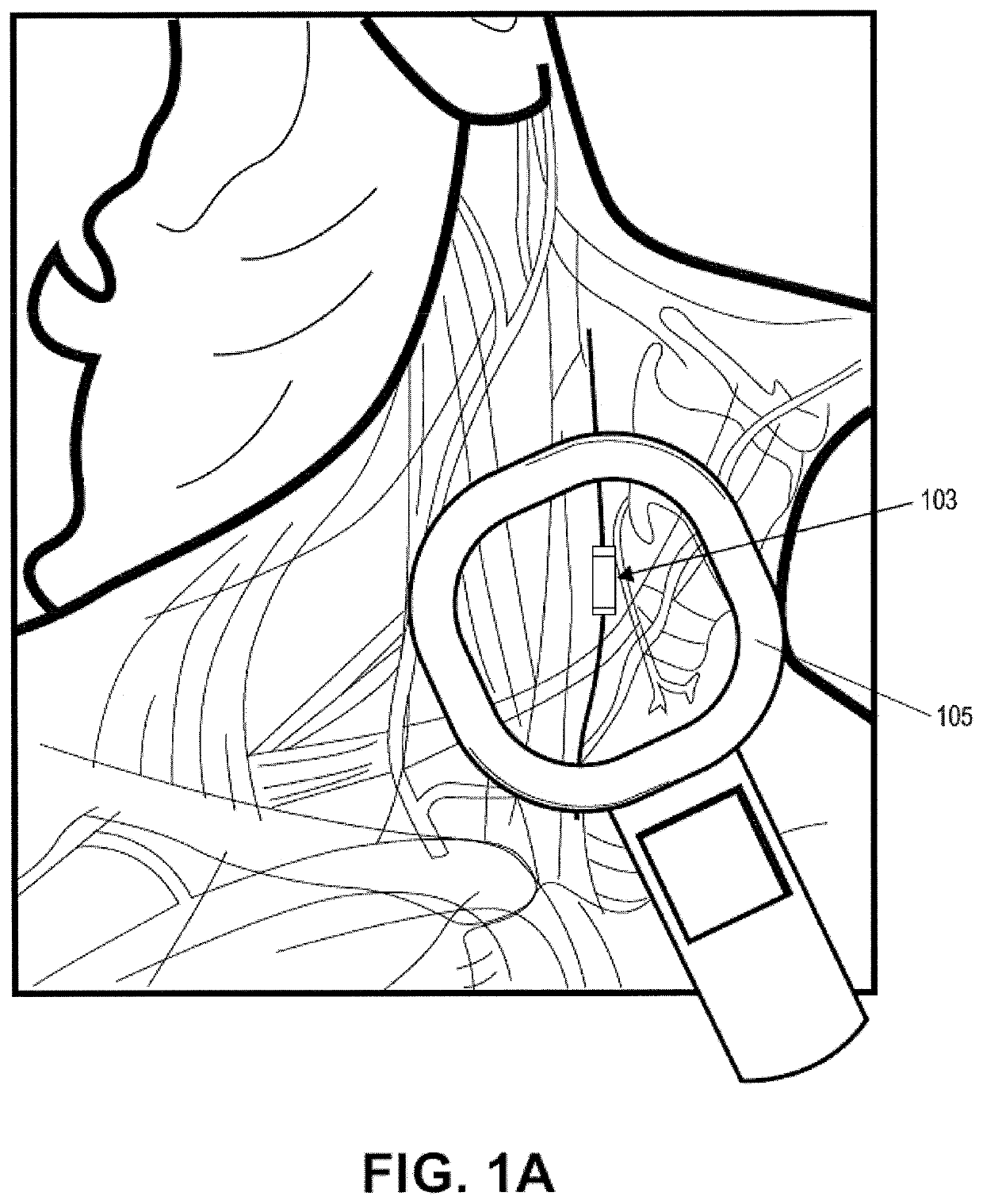
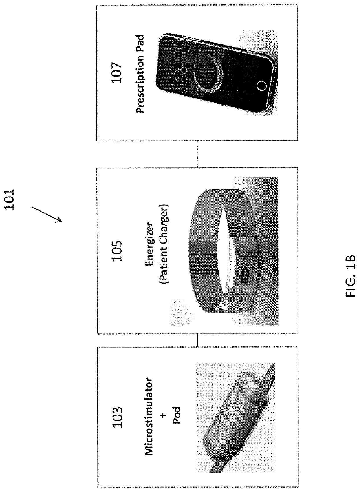
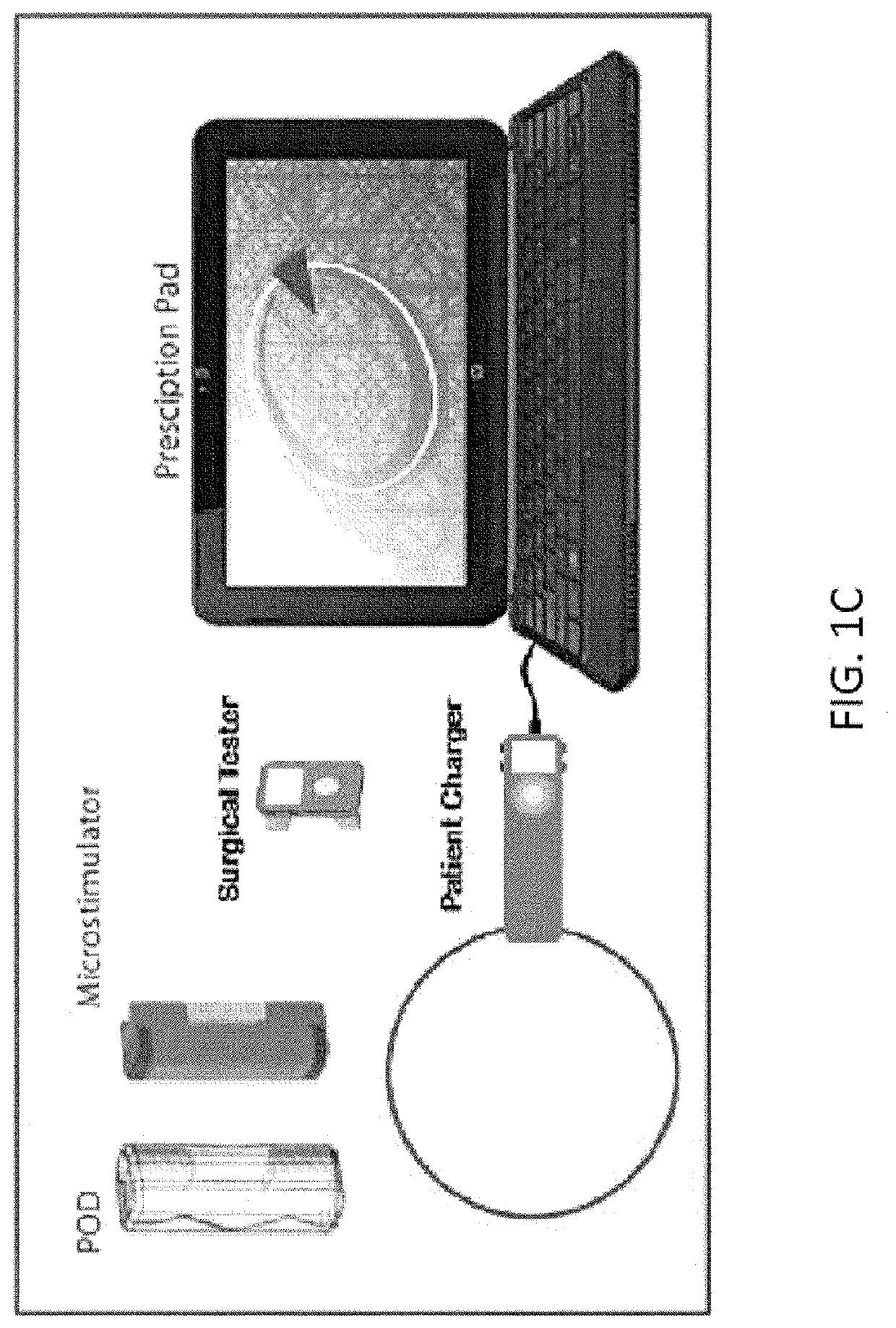
[0208]In this example, the microstimulator is a rechargeable neural stimulator that attaches to the vagus nerve and delivers current through bipolar platinum contacts against the nerve, as illustrated in FIG. 1B (103). The microstimulator battery may be charged by an external charger (“energizer”) and the energizer also functions as the communication gateway for the external controller (“prescription pad”). The Microstimulator in this example is physically housed in a rigid hermetic capsule with an attached electrode saddle. The hermetic capsule in this example is composed of an Alumina toughened Zirconia tube with metal ends brazed onto the ceramic tube. Brazing joints may use a nickel diffusion process or gold braze. The metal ends are an alloy of Titanium to reduce electrical conductivity and increase braze-ability. As shown in FIG. 23A, within the hermetic capsule is a resonator, hybrid with attached electronic parts, and batteries. The hybrid assembly is suspende...

example 2

Charger

[0295]One variation of a charger (referred to herein as an “energizer”) is described in FIGS. 36-43. In this example, the energizer for powering and programming the microstimulator attaches around the patient's neck like a necklace or collar. FIG. 36A shows one variation of the energizer around the subject's neck. In this example, about an average of 20 seconds of charging will be required per day for NCAP treatment using an implant as described above, and it is recommended that the patient charges at least every week (e.g., for approximately 20*7=3 minutes), even though it is possible not to charge for up to a month requiring a 20-30 minutes to charge.

[0296]In this example, the Energizer consists of 4 components: Coil and Magnetic Coil Connector Assembly, Electronic Module, Battery Module, and Acoustic Module.

[0297]FIG. 37 shows one variation of a coil and magnetic connector assembly. In this example, the Coil is 13 Turns of a bundle of 26 gauge magnet wire (N conductors) sp...

example

[0344]Rats were anesthetized and maintained under anesthesia with isoflurane (1-3%). A bipolar cuff electrode was placed on the left cervical vagus nerve and electrocardiogram (ECG) needle electrodes were placed subcutaneously. An electroneurogram (ENG; 20 kHz, 50,000× gain) was recorded, and an ECG (1.625 kHz, 1000× gain) was recorded with Biopac MP150 running under Acqknowledge v.4. The left vagus nerve was stimulated at 1 mA, 250 μs pulsewidth, 10 Hz for 30 seconds or with a sham stimulation (0 mA). The rats were injected intraperitoneally (IP) with lipopolysaccharide (LPS; 1 mL of a LPS solution at a concentration of 1.5 mg / mL). The data was imported into Labchart v.7 (ADInstrument) for analysis. Spectral analysis (power) of ENG and ECG data between 0-65 Hz was performed.

[0345]FIGS. 51-54I illustrate various ENGs taken of rat vagus nerves and concurrently recorded ECGs. The ENG cover a baseline period before VNS or LPS challenge, a period after VNS, and a period after LPS challe...


PUM


Abstract

Devices, systems and methods for the treatment of chronic inflammatory disorders that include an implantable microstimulator and an external charger / controller wherein the microstimulator is configured to operate using closed-loop feedback. The feedback for the microstimulator can be electrical activity of the vagus nerve and / or heart sensed by the microstimulator. The feedback can be used to modulate the stimulation duration, intensity, frequency, on-time and off-time.

Description

CROSS REFERENCE TO RELATED APPLICATIONS[0001]This application claims priority to U.S. Provisional Application No. 61 / 810,690, filed Apr. 10, 2013, which is herein incorporated by reference in its entirety.[0002]Some variations of the methods and apparatuses described in this patent application may be related to the following U.S. patent applications: U.S. patent publication no. US-2010-0125304, filed on Nov. 17, 2009 and titled, “DEVICES AND METHODS FOR OPTIMIZING ELECTRODE PLACEMENT FOR ANTI-INFLAMATORY STIMULATION”; U.S. patent publication no. US-2011-0054569, filed on Sep. 1, 2010 and titled, “PRESCRIPTION PAD FOR TREATMENT OF INFLAMMATORY DISORDERS”; U.S. patent publication no. US-2011-0106208, filed on Nov. 1, 2010and titled, “MODULATION OF THE CHOLINERGIC ANTI-INFLAMMATORY PATHWAY TO TREAT PAIN OR ADDICTION”; U.S. patent publication no. US-2011-0190849, filed on Dec. 23, 2010 and titled, “NEURAL STIMULATION DEVICES AND SYSTEMS FOR TREATMENT OF CHRONIC INFLAMMATION”; U.S. paten...

Claims


Application Information

Patent Timeline
Patent Type & Authority Patents(United States)
IPC IPC(8): A61B5/349A61N1/05A61N1/36A61N1/37A61N1/378A61B5/00A61N1/372A61N1/375
CPCA61B5/349A61B5/4047A61B5/4836A61B5/6877A61N1/0551A61N1/36053A61N1/36139A61N1/3702A61N1/3756A61N1/3787A61N1/37205
Inventor LEVINE, JACOB A.FALTYS, MICHAEL A.
Owner SETPOINT MEDICAL CORP
Learn More