Method and device for detecting circulating tumor cell
a tumor cell and detection method technology, applied in the field of biological and medical detection, can solve the problems of bringing a great technical challenge for detection, unable to fully and accurately reflect the current status of tumor focus, and the risk of metastases, so as to achieve the effect of effective and rapid detection of the degree of malignancy of free ctcs
- Summary
- Abstract
- Description
- Claims
- Application Information
AI Technical Summary
Benefits of technology
Problems solved by technology
Method used
Image
Examples
example 1
[0202]Detection of Glucose Analogue Uptake for a Very Small Number of Cells
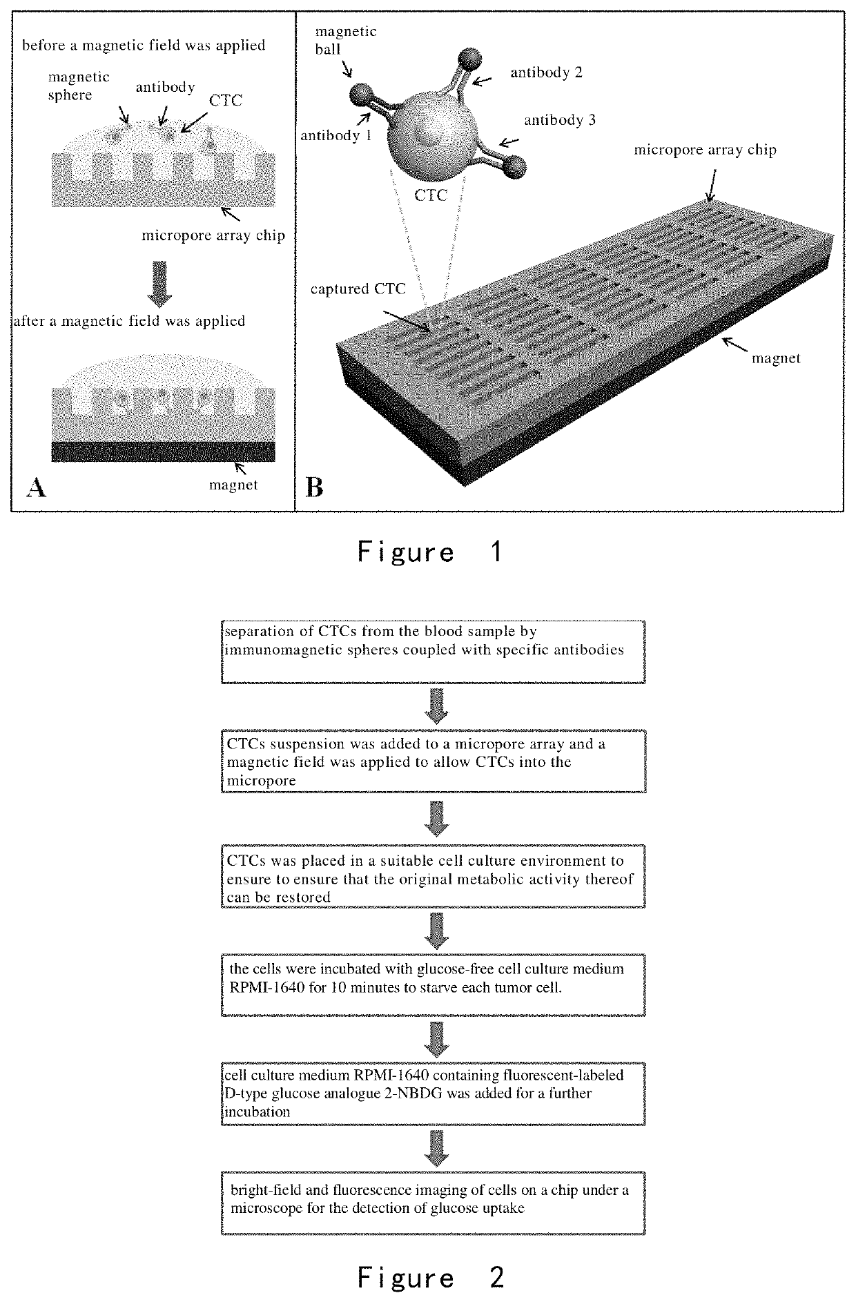
[0203]A microwell array PDMS chip was provided, and its structure is shown in FIG. 1. 30 lung cancer cells A549, H1650, and H1975 were obtained respectively, wherein the surface of each cell was combined with immunized magnetic beads, and the cells were suspended in approximately 200 microliters of PBS, respectively and placed in the microwell array PDMS chip (FIG. 1) to ensure each of them enter and fix in the microwells by manipulating the magnetic field.
[0204]As shown in FIG. 2, the cells were incubated with glucose-free cell culture medium RPMI-1640 for 10 minutes to starve each tumor cell. Then, cell culture medium RPMI-1640 containing 2-NBDG (the concentration was 0.3 mM) was added and the cells were incubated for another 20 minutes. After the incubation, the cells were washed completely and repeatedly with PBS at 4° C. on ice, and the cells were fixed in the microwells by applying a magnetic field. Fin...
example 2
[0207]Detection of Glucose Analog Uptake of CTC in Peripheral Blood
[0208]In this example, the method comprises the following steps:
[0209](1) For 2 ml of peripheral blood samples from lung cancer patients (volunteers), the upper platelet-rich plasma was first removed by low speed centrifugation (200 g) for 5 minutes, and the remaining cells were resuspended in 2 ml with Hank's balanced salt solution (HBSS), and a group of biotin-labeled antibodies (targeted antigens were EpCAM, EGFR, HER2, MUC1, respectively, with a final concentration of 1 μg / mL for each antibody) was added and co-incubated for 1 hour. The excess antibodies were centrifuged (300 g, 5 minutes) and removed, and then streptavidin-labeled magnetic balls (0.8 μm) were added for co-incubation for 30 min, excess magnetic balls were removed by centrifugation (300 g, 5 min), and the cells were resuspended in 5 ml of HBSS to form the pretreated sample.
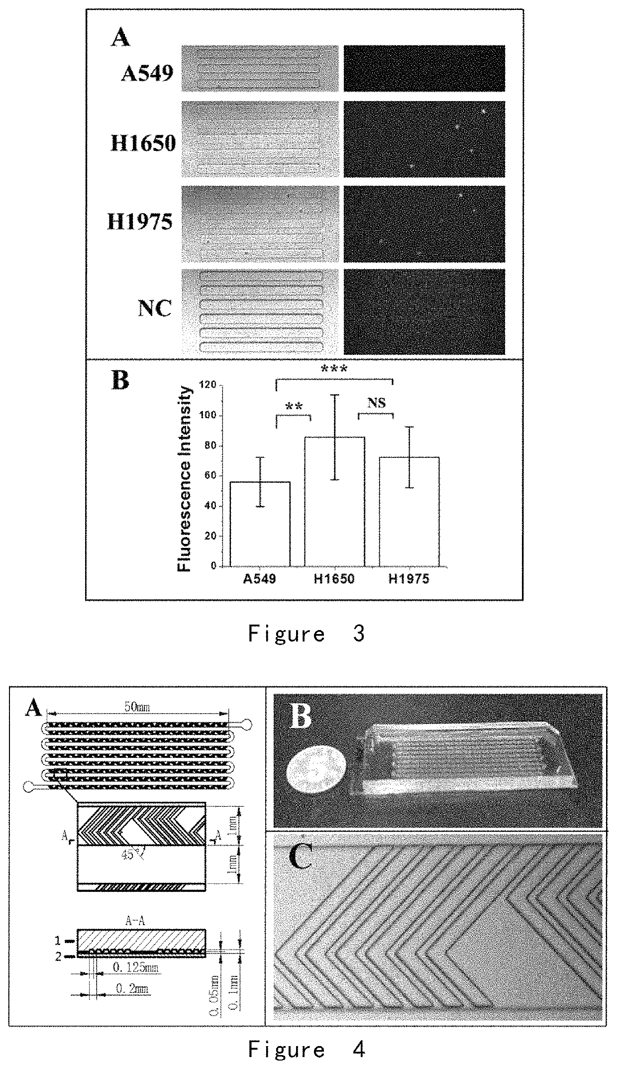
[0210](2) A fishbone chip was provided, with a structure shown in FIG. 4.
[0...
example 3
[0220]Example 2 was repeated, wherein the peripheral blood sample used was the same batch of peripheral blood samples from the same lung cancer patient (volunteer). The difference is that in step (5), the upper conditioned medium in the culture dish of lung cancer cell H1650 and the upper conditioned medium in the lung fibroblast culture dish were combined in a ratio of 1:1 to form a new conditioned medium for CTC culturing in microchip.
[0221]As a result, similar results as in Example 2 were obtained, and significant fluorescent signals were detected in 6 CTCs.
PUM
| Property | Measurement | Unit |
|---|---|---|
| length | aaaaa | aaaaa |
| length | aaaaa | aaaaa |
| width | aaaaa | aaaaa |
Abstract
Description
Claims
Application Information
 - R&D
- Intellectual Property
- Life Sciences
- Materials
- Tech Scout
- Unparalleled Data Quality
- Higher Quality Content
- 60% Fewer Hallucinations
Browse by: Latest US Patents, China's latest patents, Technical Efficacy Thesaurus, Application Domain, Technology Topic, Popular Technical Reports.
© 2025 PatSnap. All rights reserved.Legal|Privacy policy|Modern Slavery Act Transparency Statement|Sitemap|About US| Contact US: help@patsnap.com


