
Particle size purification method and devices

a technology of particle size and purification method, which is applied in the field of biological fluid screening methods, can solve the problems of high background reading distortion, high cost of reagents, and the necessity that exosomes contain antigens, and achieve the effect of providing an analysis quickly

Active Publication Date: 2021-11-02
UNIV OF NOTRE DAME DU LAC
View PDF10 Cites 0 Cited by
  • Summary
  • Abstract
  • Description
  • Claims
  • Application Information

AI Technical Summary

Benefits of technology

[0017]In another aspect, the invention provides an exosome purification process / tool. The advantages of these processes / tools include that it provides an analysis quickly, requires a relatively small amount of sample biological fluid / test material, and may be used with a variety of body fluids, including urine, blood, serum, plasma, saliva, mucus, and other biological fluids.

Problems solved by technology

The selection of particular particles of interest from a sample, such as from a biological sample, presents several challenges related to yield and purity.
However, the current commercial kits / reagents for exosome purification are based on non-specific precipitation using polymers, size exclusion chromatography or antibody-based affinity purification.
These approaches suffer various disadvantages, including high background readings that distort results and mask detection of the diagnostic molecule of interest in a sample (soluble protein / component contamination in the final preparation) or in the case of an antibody-based approach, the cost of the reagents and the necessity that the exosome contain the antigen recognized by the antibody.

Method used


Image

  • Particle size purification method and devices
  • Particle size purification method and devices
  • Particle size purification method and devices

Examples


example 1

er Filter Unit

[0049]The present example details a specific configuration of the multimember filter unit that is included in the particle size separation device. The particle size separation device is referred to herein as an “ExoPen”.

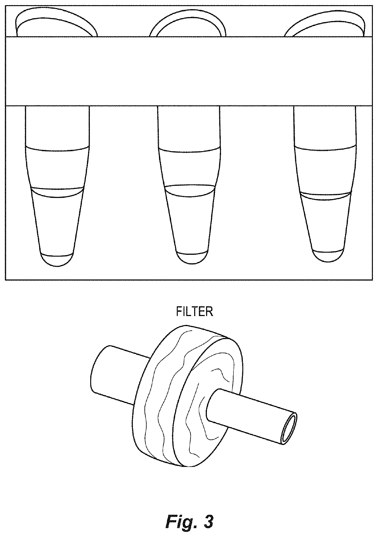
[0050]The multimember filter unit comprises multiple membranes, these membranes having very small pore sizes, and are configured as part of the present separation device in a series of membranes having decreasing pore sizes. For example, a membrane providing a 80 μm filter, a membrane providing a 10 μm filter and a membrane providing a 0.2 μm filter, in series, will be assembled as part of one configuration. In addition, to make this multi-membrane unit, the 0.2 μm filter unit is separated and additional filters are added on top of the 0.2 μm filter membrane, these filter membranes being divided spatially by thin washers (see FIG. 3). The whole multi-filter construct is glued together to produce a multi-membrane filter unit (FIG. 4).

[0051]The configurat...

example 2

Exosome Purification

[0054]The present example presents a description of a method of isolating and / or providing a purified preparation of exosomes from a biological fluid using the multi-filter unit described above, such as in an ExoPen construct.

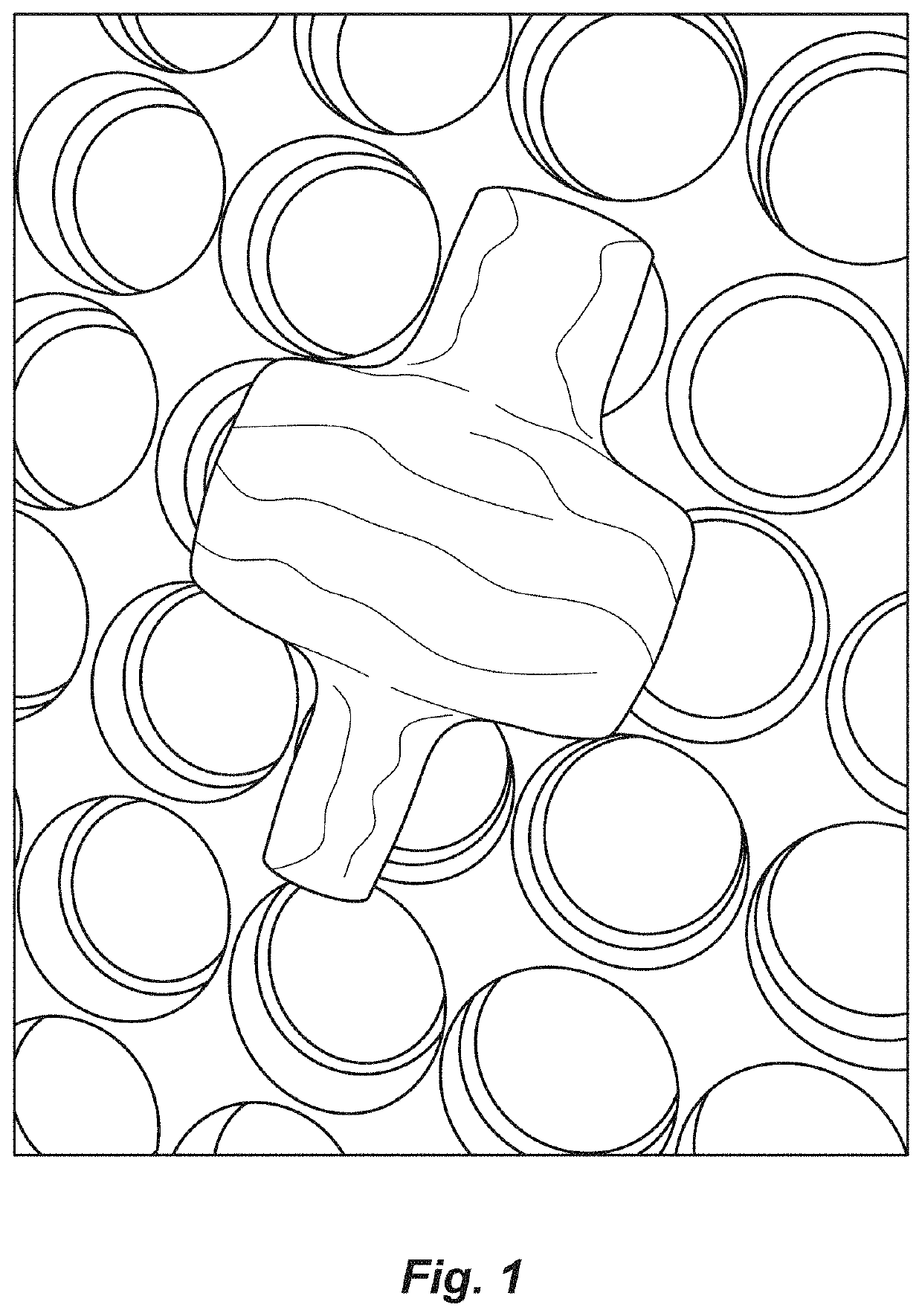
[0055]A biological fluid, such as a body fluid (serum, blood, plasma, ascites fluid) is passed through a multi-membrane filter unit (See filter depicted at FIG. 1). Any remaining biological fluid contained within the filter unit is collected by adding PBST to the filter unit and is collected as the PBST buffer is being pushed into the membrane unit.

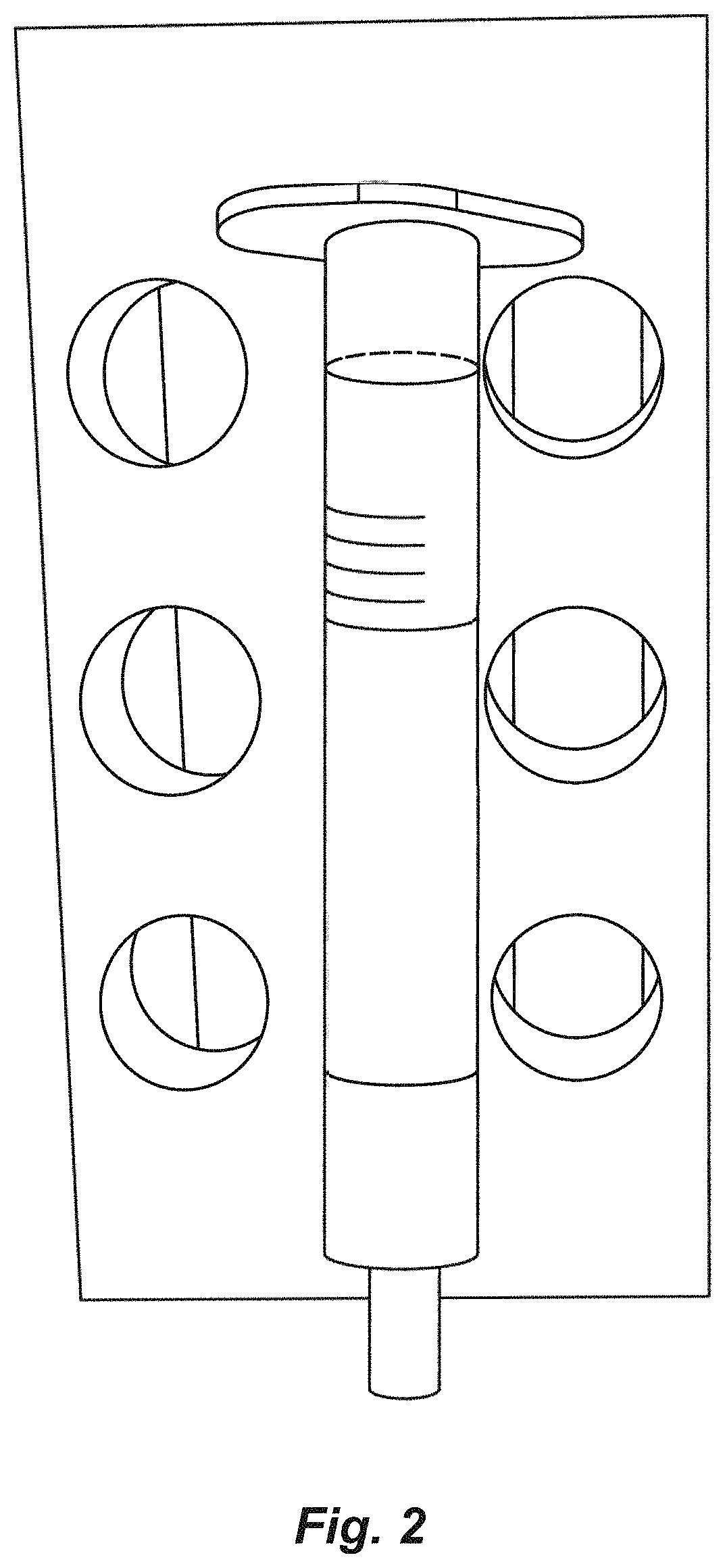
[0056]A column (FIG. 2) was prepared using a plastic hollow cylinder with a mesh at one end that was used to hold a resin within the cylinder. GE CAPTO Core 700 beads are loaded into the column and equilibrated with 10 ml of washing buffer (PBST).

[0057]The filtered body fluid, which contains free protein and vesicles smaller then 200 nm, was loaded onto the equilibrated column, and then washing bu...

example 4

and Methods

[0060]The present example provides the materials and methods (flow chart) by which a sample may be processed to isolate by particular size, the various components in the sample. In this example, a human serum is the fluid examined. The particles being isolated are exosomes.

[0061]FIG. 5 presents a list of the types of methods employed in the analysis of a sample in assessing exosome purification efficiency employing both an ExoPen-based or EXOQUICK product-based exosome purification of a 10 human serum samples obtained from patients with a confirmed tuberculosis disease.

[0062]FIG. 6 provides a flow-chart of the workflow procedure for processing the human serum samples via either the ExoPen method (Tube 1) or the EXOQUICK product method (Tube 2).

[0063]EXOQUICK product reagent is added to serum at a 4:1 ratio (25 μl EXOQUICK product per 100 μl of serum). The sample is mixed and kept on ice for a minimal of 1 hour but can be kept at 4° C. overnight. The sample containing the ...


PUM

PropertyMeasurementUnit
sizeaaaaaaaaaa
sizeaaaaaaaaaa
sizeaaaaaaaaaa

Abstract

A particle separation multi-membrane matrix device and method are provided. The particles isolated may comprise nano-scale particles, such extracellular membrane vesicles, having a size of about 50 to about 150 nm. The vesicles are released by many different cell types, and may be efficiently isolated at high yield and purity according to the present methods from various body fluids (e.g., blood, saliva, breast milk, serum, plasma, ascites fluid, etc.). Such isolated exosome preparations may include biomarkers, such as disease biomarkers (diagnostic markers) for various disease (early stage and late stage cancers, neurological disorders (Parkinson disease, Alzheimer disease), diabetes, pancreatic diseases, renal failure, infectious diseases (HIV, tuberculosis, malaria, hepatitis)). The present methods and devices may be used to detect and monitor animals (human, livestock, companion animal) for infectious diseases, such as tuberculosis and other diseases. The methods and devices require minimal sample material (10 μl), are rapid, economical, yield highly enriched small molecule (eg, exosomes) preparations, and do not require electricity.

Description

CROSS-REFERENCE TO RELATED APPLICATIONS[0001]The present application claims priority to U.S. Ser. No. 62 / 252,641, filed Nov. 9, 2015, the entire contents of which are specifically incorporated herein by reference in its entirety.STATEMENT REGARDING FEDERALLY SPONSORED RESEARCH OR DEVELOPMENT[0002]NoneFIELD OF THE INVENTION[0003]This invention relates to the field of methods for screening biological fluids for biomarkers, such as biomarkers for disease, as well as devices for screening biological fluids and isolating materials from a biological fluid, such as exosomes.BACKGROUND OF THE INVENTION[0004]The selection of particular particles of interest from a sample, such as from a biological sample, presents several challenges related to yield and purity. Virtually all existing techniques require some form of sophisticated laboratory equipment, electricity source, and a more than a minimal volume of a sample to provide.[0005]Exosomes are nano-scale extracellular membrane vesicles of 50...

Claims


Application Information

Patent Timeline
Patent Type & Authority Patents(United States)
IPC IPC(8): G01N1/40B01D61/14B01D61/18B01D63/08G01N33/49
CPCG01N1/4077B01D61/142B01D61/147B01D61/18B01D63/082G01N33/491B01D2313/143B01D2313/146B01D2319/025B01D2319/06G01N2001/4088Y02A50/30B01D61/146
Inventor SCHOREY, JEFFREY S.CHENG, YONG
Owner UNIV OF NOTRE DAME DU LAC
Learn More