Detection of cells in a liquid sample

a liquid sample and cell technology, applied in the field of detection of cells in liquid samples, can solve the problems of milk unsuitable for human consumption, harmful to consumers, mastitis in cows, etc., and achieve the effects of reducing interfering fluorescence signals, simple and rapid method, and efficient entry of dyes

Active Publication Date: 2022-08-16
PERKINELMER U S LLC
View PDF41 Cites 0 Cited by
  • Summary
  • Abstract
  • Description
  • Claims
  • Application Information

AI Technical Summary

Benefits of technology

[0148]The mixture of the staining composition and the sample is incubated in step c) under conditions that renders the cells permeable to the nucleic acid dye. In a preferred embodiment, the incubation is performed at a temperature from about 45 to about 95° C., such as from about 47 to about 90° C., from about 50 to about 85° C., from about 52 to about 80° C., from about 57 to about 75° C., from about 60 to about 70° C., from 61 about to about 69° C. or from about 62 to about 68° C. As already explained in the above, pH during the incubation step will be influenced by the increased temperature and will therefore differ from the pH in the staining composition. Preferably, pH during the incubation step c) is from about 8 to about 11.5, such as from about 8 to about 11, from about 8 to about 10.6, from about 8.5 to about 10.6. The incubation in step c) is suitably performed for less than 10 minutes, such as for less than 9, such as less than 8, 7, 6, 4, 3 or 2 minutes, and preferably for approximately 1 minute. The short incubation time required in step c) while still obtaining a reliable cell count, is a major advantage of the present invention.
[0150]After the incubation step c) and optionally the sonication step d) the stained cells are counted by using fluorescence detecting instrument set to detect the fluorescence of the dimeric nucleic acid dye used. The skilled person is aware of different methods to count stained cells in solution. In a preferred aspect of the present invention, the stained cells are counted by using a fluorescence detecting instrument, such as a flow cytometer, a fluorescence microscope or fluorescent imaging system, a fluorometer or a fluorescence plate reader. The detection of the fluorescence from the labeled cells, such as bacteria, is preferably performed using a flow cytometer (also referred to as FCM).
[0151]In one embodiment, somatic and bacterial cells are counted simultaneously.
[0152]In a highly preferred embodiment of the invention, a method of staining (and counting) bacteria in raw milk is performed as follows: Savinase® (or Alcalase®) and GelGreen™ are added to a Tris-saline solution (pH 10.6), further referred to as the staining composition; this staining composition is preheated to a temperature of from about 45 to about 95° C., such as from about 47 to about 90° C., from about 50 to about 85° C., from about 52 to about 80° C., from about 57 to about 75° C., from about 60 to about 70° C., from 61 about to about 69° C. or preferably from about 62 to about 68° C.; a small volume of raw milk sample is preheated just long enough for the sample to reach a temperature of from about 45 to about 95° C., such as from about 47 to about 90° C., from about 50 to about 85° C., from about 52 to about 80° C., from about 57 to about 75° C., from about 60 to about 70° C., from 61 about to about 69° C. or preferably from about 62 to about 68° C.; the preheated sample is preferably diluted about 1:10 in the preheated staining composition; the mixture of the sample with the staining composition is optionally sonicated for approximately 5 to 10 seconds by either direct placement of the sonicator probe into the sample, or indirectly by placing the appropriate sonicator probe against the cup in which the sample is held; the sample with the staining composition is incubated (preferably in a mixer cup) at a temperature of from about 45 to about 95° C., such as from about 47 to about 90° C., from about 50 to about 85° C., from about 52 to about 80° C., from about 57 to about 75° C., from about 60 to about 70° C., from 61 about to about 69° C. or preferably from about 62 to about 68° C., up to 1 to 2 min; the sample is then optionally again sonicated for approximately 5 to 10 seconds, again either directly or indirectly; and finally the mixture (or a representative part thereof) is analyzed on the flow cytometer and the fluorescent signals from the somatic cells and / or bacteria are detected and a count per unit volume is determined. This entire procedure from taking the sample to read-out takes less than 10 minutes, and preferably less than 9, 8, 7, 6, 5, 4, 3, 2, or 1 min. In an alternative method, the milk sample itself is not preheated before mixing, but becomes heated when mixed with a preheated staining composition to a temperature that allows optimal entry of the dye into the cells / microorganisms.
[0153]In one preferred aspect, said protease and said dimeric nucleic acid dye are mixed in a Tris-buffered saline to form said staining composition. And in yet another preferred embodiment of the present invention, a method is provided according to the invention wherein said staining composition is heated to from about 45 to about 95° C., such as from about 47 to about 90° C., from about 50 to about 85° C., from about 52 to about 80° C., from about 57 to about 75° C., from about 60 to about 70° C., from 61 about to about 69° C. or preferably from about 62 to about 68° C., before it is mixed with said sample. In concert therewith, preferably said sample is heated to from about 45 to about 95° C., such as from about 47 to about 90° C., from about 50 to about 85° C., from about 52 to about 80° C., from about 57 to about 75° C., from about 60 to about 70° C., from 61 about to about 69° C. or preferably from about 62 to about 68° C., when it is mixed with said staining composition. The sample may also be preheated directly before mixing with the staining composition. Depending on the type of sample the skilled person is able to decide whether to preheat or to use a sample at its original temperature before mixing (milk is generally stored in a cold place, or when directly derived from the source such as a cow, the milk may have a temperature that is above storage temperature, but below incubation temperature). When the sample and staining composition are mixed, the incubation takes place at the preferred and optimal temperatures to obtain the most efficient entry of the dye into the cells. Alternatively, the sample is not preheated (and has a cold storage temperature, for instance), but reaches an optimal temperature for dye entry when it is mixed with the preheated staining composition.
[0154]In a highly preferred embodiment, the present invention relates to a method of counting cells, such as somatic and / or bacterial cells in milk, such as cow milk, said method comprising the steps of:

Problems solved by technology

In agriculture, the presence of bacteria such as Staphylococcus aureus, may cause mastitis in cows and may be harmful for consumers when present in milk.
The art teaches a variety of methods to determine the number of somatic cells in milk, which in turn is clearly an indication that the milk is unsuitable for human consumption.
This mixture causes lysis of the cells, degrades and solubilizes the protein particles and cell debris and stains the bacteria in the sample.
The disadvantage of this method lies in the number of agents that are used (including an ion-chelating agent and a detergent).
The use of toxic agents results in cumbersome waste handling and higher costs per sample.
The disadvantage of this method is that it is time consuming as it requires centrifugation and a dye is used that depends on the viability of the cells and can therefore not detect all cells, including certain living cells.
This method has the disadvantage that the incubation period between bacteria and fluorogenic substrate takes at least 7 hours, which is too long for rapid processing of milk products.
Many of these nucleic acid amplification / detection procedures take time and sometimes require the breakdown of the bacteria and extraction and / or precipitation of the genetic material, which makes these procedures generally complex and cumbersome.
The disadvantage of using this method is the staining procedure and the use of specific antibodies against specific bacteria (that might miss bacteria not bound to such antibodies).
Moreover, the relative high costs that come with using (recombinant) antibodies is an additional disadvantage of that procedure.
The disadvantage in that particular method lies in the complicated de-proteinization and purification method that takes time.
This method has the disadvantage of using multiple and costly dyes, and time: the different steps add up to a procedure that will last between 1 and 48 hours.
The disadvantage of this method is that Gunasekera et al, discloses an additional centrifugation step, which takes time and is cumbersome in rapid detection systems.
This is still a relatively long procedure.
None of the above methods known in the art teach a procedure that is fast, requires only a few steps, that is reliable, in-expensive and safe.
Many of the listed methods are either slow, use toxic reagents, are cumbersome and / or expensive, which makes them less suitable for large production processes and high-throughput screenings.

Method used


Image

  • Detection of cells in a liquid sample
  • Detection of cells in a liquid sample
  • Detection of cells in a liquid sample

Examples


example 1

Validation of GelGreen™ Staining of Bacteria

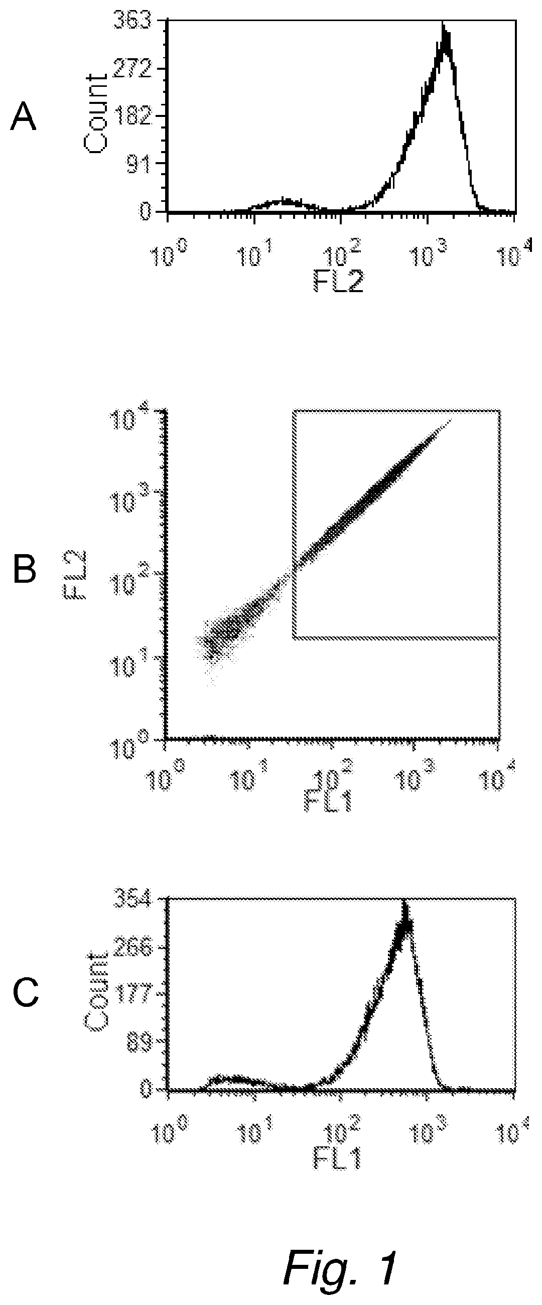
[0198]First, an experiment was performed to confirm that bacterial counts obtained on the flow cytometer using GelGreen™ did correlate with traditional plate counts. For this, the Petrifilm method was selected (Standard Methods for Examination of Dairy Products 17th Edition 2004. Edited by H. M. Wehr and J. R. Frank. American Public Health Association, 800 I Street, NW, Washington D.C. 200001. Chapter 6.040), rather than a poured agar plate method, because of ease of use. Escherichia coil (E. coli) cultures were grown at 36° C. in Trypticase Soy Broth (TSB) in a culture tube. A staining composition was prepared as follows: The nucleic acid dye GelGreen™ (from Biotium) was diluted to 1× from the 10,000× commercially available stock, into Tris-buffered saline (100 mM Trizma base, 100 mM Trizma HCl, 150 mM NaCl, pH 8.5), which was preheated to 50° C. The used dilution of 1:10,000 of the GelGreen™ is in accordance with the manufacturer's recom...

example 2

Staining of E. coli in Raw Milk Treated with GelGreen™

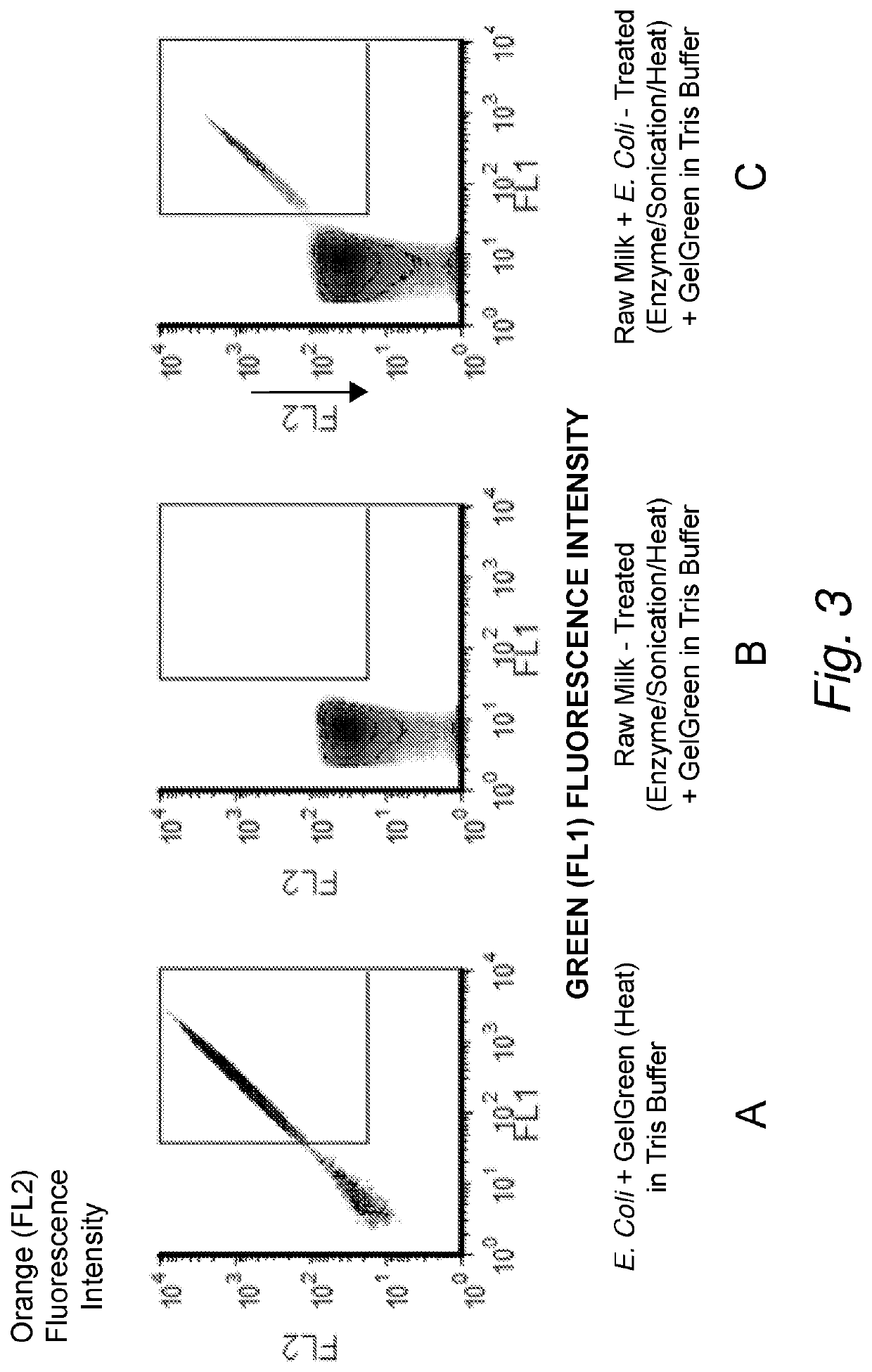
[0199]To show where the bacteria in the FL1 and FL2 signal plots can be expected, bacteria were first counted in the staining composition without adding any milk. A culture of E. coli were grown at 36° C. in Trypticase Soy Broth (TSB) in a culture tube. The nucleic acid dye GelGreen™ was diluted into Tris-buffered saline, at approximately pH 8.5, which was preheated to 50° C. The E. coli culture was removed from the 36° C. incubator and a small volume was diluted 1:100 into the preheated staining composition and incubated for approximately 4.5 min at 50° C. The stained E. coli was analyzed on the flow cytometer and the FL1 and FL2 fluorescence channels were used to identify the stained bacteria (FIG. 3A).

[0200]To show how the FL1, FL2 signals look like in un-spiked raw milk, low bacterial count raw milk was analyzed. The locally obtained raw milk (generally from a farm) has a very low bacteria count ranging from 1000 to 50,000 CF...

example 3

Validation of Bacterial Flow Cytometry Counts in Raw Milk

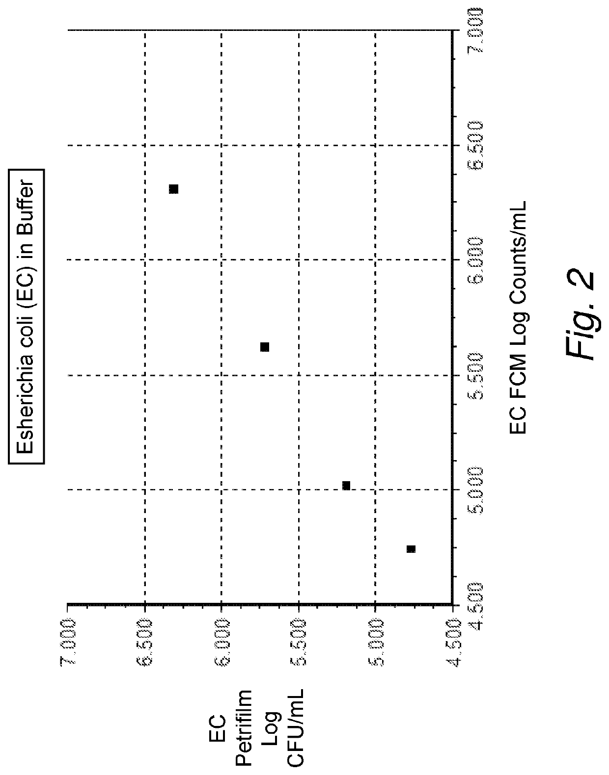
[0202]A selection of gram negative and gram positive bacteria were chosen to be placed into raw milk and processed and analyzed following the GelGreen™ raw milk treatment staining protocol as disclosed in example 2. Results are shown in FIG. 4. The gram positive organisms selected were Listeria monocytogenes (Lm), Staphylococcus aureus (Sa), and Bacillus cereus (Bc). The gram negative organisms selected were Pseudomonas aeruginosa (Pa) and Escherichia coil (Ec). Different concentrations of each culture were placed into a sample of raw milk and kept at 4° C. until processed. Each sample of raw milk with an individual bacterial culture was processed and analyzed following the GelGreen™ protocol as described in example 2. Each sample of raw milk with an individual bacterial culture was appropriately diluted in Butterfield's PBS and 1 mL was plated on Petrifilms in duplicate and CFU / mL were obtained. The resulting Log FCM counts p...


PUM

PropertyMeasurementUnit
temperatureaaaaaaaaaa

Abstract

The invention relates to a method for counting cells, such as bacteria and / or somatic cells in liquid samples, such as in dairy products, preferably raw milk. Disclosed is a method comprising a combination of steps that apply dimeric nucleic acid dyes that normally do not penetrate cells (=cell-impermeant dyes), which are rendered cell-permeant by using the right combination of pH, buffer and temperature.

Description

[0001]This application is the U.S. national phase of International Application No. PCT / M2017 / 000364 filed Mar. 17, 2017 which designated the U.S. and claims priority to European patent application Serial No. EP18160837.7 filed 17 Mar. 2016 the entire contents of each of which are hereby incorporated by reference.RELATED APPLICATIONS AND INCORPORATION BY REFERENCE[0002]This application claims benefit of European patent application Serial No. EP16160837.7 filed 17 Mar. 2016.[0003]The foregoing applications, and all documents cited therein or during their prosecution (“appln cited documents”) and all documents cited or referenced in the appln cited documents, and all documents cited or referenced herein (“herein cited documents”), and all documents cited or referenced in herein cited documents, together with any manufacturer's instructions, descriptions, product specifications, and product sheets for any products mentioned herein or in any document incorporated by reference herein, are...

Claims


Application Information

Patent Timeline
Patent Type & Authority Patents(United States)
IPC IPC(8): G01N1/30C12Q1/04A01B5/02C12M1/34B01L3/00C09B11/28C09B23/04C12Q1/68G01N21/64
CPCG01N1/30A01B5/02B01L3/5023B01L3/50273C09B11/28C09B23/04C12M1/34C12Q1/04C12Q1/68G01N21/6486
Inventor EMOND, PIERRE LAURENTPERLMUTTER, NANCY GAILKREIDER, JAMES WILLIS
Owner PERKINELMER U S LLC
Why Eureka
Try Eureka